

The impact of parental contact upon cortical noxious-related activity in human neonates
- PMID: 32965725
- PMCID: PMC8436758
- DOI: 10.1002/ejp.1656
The impact of parental contact upon cortical noxious-related activity in human neonates
Abstract
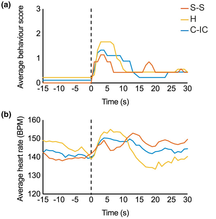
Background: Neonates display strong behavioural, physiological and cortical responses to tissue-damaging procedures. Parental contact can successfully regulate general behavioural and physiological reactivity of the infant, but it is not known whether it can influence noxious-related activity in the brain. Brain activity is highly dependent upon maternal presence in animal models, and therefore this could be an important contextual factor in human infant pain-related brain activity.
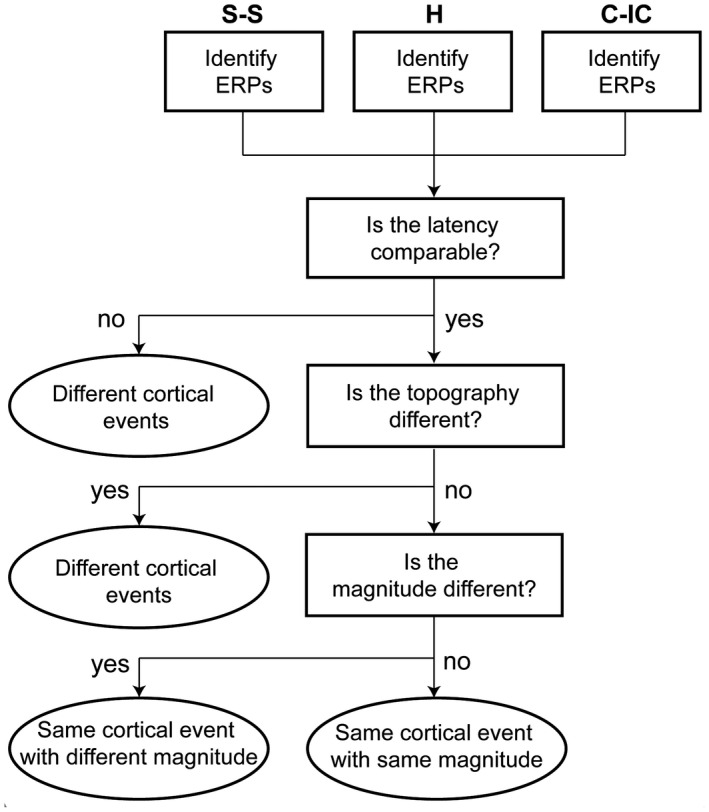
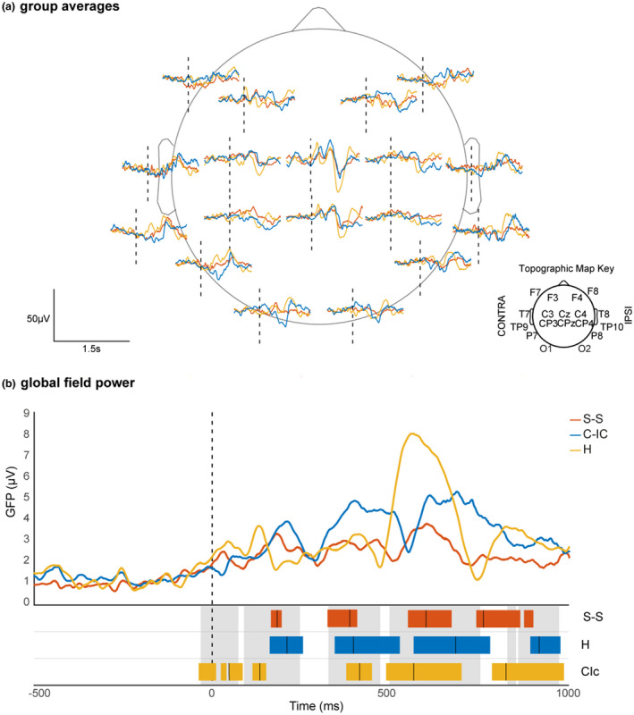
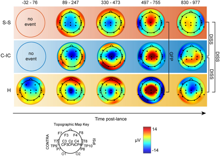
Methods: Global topographic analysis was used to identify the presence and inter-group differences in noxious-related activity in three separate parental contexts. EEG was recorded during a clinically required heel lance in three age and sex-matched groups of neonates (a) while held by a parent in skin-to-skin (n = 9), (b) while held by a parent with clothing (n = 9) or (c) not held at all, but in individualized care (n = 9).
Results: The lance elicited a sequence of 4-5 event-related potentials (ERPs), including the noxious ERP (nERP), which was smallest for infants held skin-to-skin and largest for infants held with clothing (p=0.016). The nERP was then followed by additional and divergent long-latency ERPs (> 750 ms post-lance), not previously described, in each of the groups, suggesting the engagement of different higher level cortical processes depending on parental contact.
Conclusions: These results show the importance of considering contextual factors in determining infant brain activity and reveal the powerful influence of parental contact upon noxious-related activity across the developing human brain.
Significance: This observational study found that the way in which the neonatal brain processes a noxious stimulus is altered by the type of contact the infant has with their mother. Specifically, being held in skin-to-skin reduces the magnitude of noxious-related cortical activity. This work has also shown that different neural mechanisms are engaged depending on the mother/infant context, suggesting maternal contact can change how a baby's brain processes a noxious stimulus.
© 2020 The Authors. European Journal of Pain published by John Wiley & Sons Ltd on behalf of European Pain Federation - EFIC®.
Conflict of interest statement
The authors have no conflict of interest to declare.
Figures
Similar articles
-
Establishing a standardised approach for the measurement of neonatal noxious-evoked brain activity in response to an acute somatic nociceptive heel lance stimulus.Cortex. 2024 Oct;179:215-234. doi: 10.1016/j.cortex.2024.05.023. Epub 2024 Jul 27. Cortex. 2024. PMID: 39197410 Free PMC article.
-
Effect of parental touch on relieving acute procedural pain in neonates and parental anxiety (Petal): a multicentre, randomised controlled trial in the UK.Lancet Child Adolesc Health. 2024 Apr;8(4):259-269. doi: 10.1016/S2352-4642(23)00340-1. Epub 2024 Feb 16. Lancet Child Adolesc Health. 2024. PMID: 38373429 Clinical Trial.
-
The influence of skin-to-skin contact on Cortical Activity during Painful procedures in preterm infants in the neonatal intensive care unit (iCAP mini): study protocol for a randomized control trial.Trials. 2022 Jun 20;23(1):512. doi: 10.1186/s13063-022-06424-4. Trials. 2022. PMID: 35725632 Free PMC article.
-
Can Event-Related Potentials Evoked by Heel Lance Assess Pain Processing in Neonates? A Systematic Review.Children (Basel). 2021 Jan 20;8(2):58. doi: 10.3390/children8020058. Children (Basel). 2021. PMID: 33498331 Free PMC article. Review.
-
Oral morphine analgesia for preventing pain during invasive procedures in non-ventilated premature infants in hospital: the Poppi RCT.Southampton (UK): NIHR Journals Library; 2019 Aug. Southampton (UK): NIHR Journals Library; 2019 Aug. PMID: 31483590 Free Books & Documents. Review.
Cited by
-
Maternal speech decreases pain scores and increases oxytocin levels in preterm infants during painful procedures.Sci Rep. 2021 Aug 27;11(1):17301. doi: 10.1038/s41598-021-96840-4. Sci Rep. 2021. PMID: 34453088 Free PMC article.
-
Psychosocial and Neurobiological Vulnerabilities of the Hospitalized Preterm Infant and Relevant Non-pharmacological Pain Mitigation Strategies.Front Pediatr. 2021 Oct 25;9:568755. doi: 10.3389/fped.2021.568755. eCollection 2021. Front Pediatr. 2021. PMID: 34760849 Free PMC article. Review.
-
Parent-led neonatal pain management-a narrative review and update of research and practices.Front Pain Res (Lausanne). 2024 Apr 16;5:1375868. doi: 10.3389/fpain.2024.1375868. eCollection 2024. Front Pain Res (Lausanne). 2024. PMID: 38689885 Free PMC article. Review.
-
Assessment and Management of Pain in Preterm Infants: A Practice Update.Children (Basel). 2022 Feb 11;9(2):244. doi: 10.3390/children9020244. Children (Basel). 2022. PMID: 35204964 Free PMC article. Review.
-
Decoding of pain during heel lancing in human neonates with EEG signal and machine learning approach.Sci Rep. 2024 Dec 28;14(1):31244. doi: 10.1038/s41598-024-82631-0. Sci Rep. 2024. PMID: 39732802 Free PMC article.
References
-
- Allison, T., McCarthy, G., & Wood, C. C. (1992). The relationship between human long‐latency somatosensory evoked potentials recorded from the cortical surface and from the scalp. Electroencephalography and Clinical Neurophysiology/Evoked Potentials Section, 84(4), 301–314. 10.1016/0168-5597(92)90082-M - DOI - PubMed
-
- Als, H. (2009). Newborn individualized developmental care and assessment program (NIDCAP): New frontier for neonatal and perinatal medicine. Journal of Neonatal‐Perinatal Medicine, 2(3), 135–147. 10.3233/NPM-2009-0061 - DOI
Publication types
MeSH terms
Grants and funding
LinkOut - more resources
Full Text Sources
Medical
