

Gene expression of the immunoinflammatory and immunological status of obese dogs before and after weight loss
- PMID: 32966299
- PMCID: PMC7510989
- DOI: 10.1371/journal.pone.0238638
Gene expression of the immunoinflammatory and immunological status of obese dogs before and after weight loss
Abstract
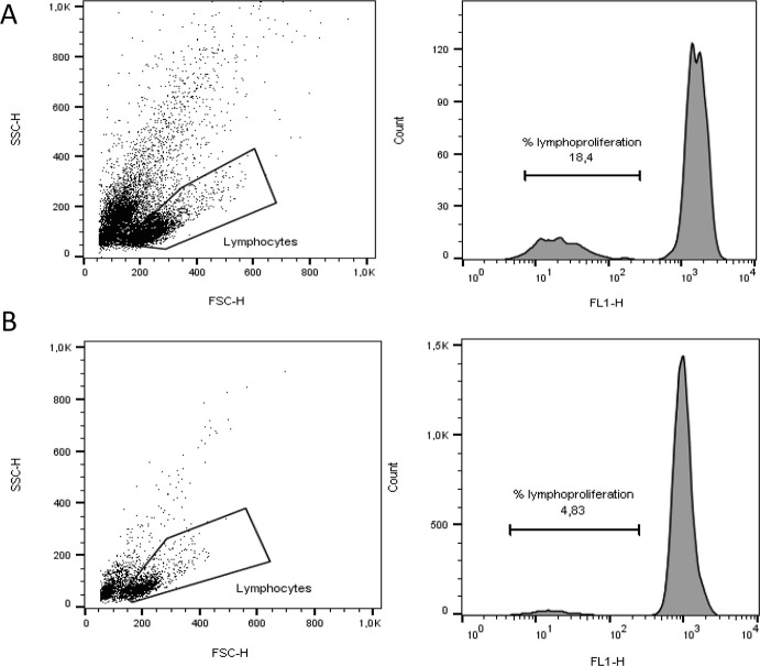
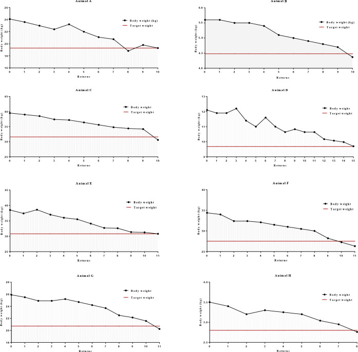
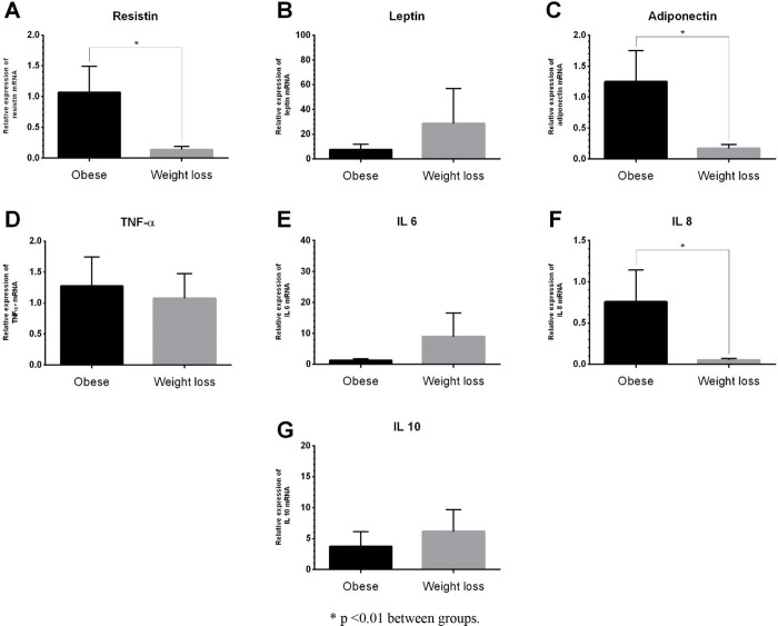
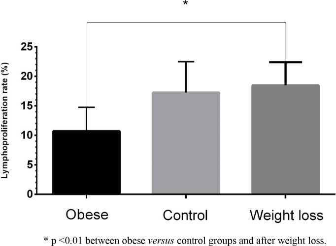
Obesity is characterized by a low degree of chronic inflammation state that, along with metabolic modifications, promotes important changes in the animal's organism. Adipose tissue actively participates in inflammation and immunity, and several defense cells of the organism may, therefore, be involved in the diversity found between obese and ideal weight individuals. Studies regarding this subject have shown immune cell changes in humans and rats, however, the literature is scarce in relation to dogs. Thus, the present study aimed to evaluate the gene expression profile of immunoinflammatory response and the lymphoproliferation of obese dogs before and after weight loss. Eight female dogs, neutered, of different breeds, aged between 1 and 8 years (4.74±3.19), obese, with body condition score (BCS) of 9 out of a 9-point scale and body composition determined by the deuterium isotope dilution method were included. The obese dogs were enrolled in a weight loss program and after losing 20% of their initial weight became a second experimental group. A third experimental group consisted of eight female dogs, neutered, aged between 1 and 8 years (3.11±0.78) and with ideal BCS (5 out of a 9-point scale). Gene expression of immunoinflammatory cytokines (resistin, leptin, adiponectin, TNF-α, IL-6, IL-8, and IL-10) was assessed by qRT-PCR and immunity was assessed by lymphoproliferative response using the flow cytometry technique. The data that presented normal distribution was evaluated by analysis of variance by the PROC MIXED of the SAS and when differences were detected, these were compared by the Tukey test. Regarding the gene expression data, the procedure PROC GLIMMIX was adopted and the methodology of generalized linear model was used, in which the Gama distribution proved to be adequate. Values of p<0.05 were considered significant. The mean weight loss period of the animals included in the study was 194.25 ± 28.31 days and the mean weekly weight loss rate was 1.02 ± 0.82%. The average fat mass, both in percentage (P<0.001) and in kilograms (P = 0.012), was higher in the obese group (40.88%; 8.91kg), returning to normal and without difference between the control group (19.16%; 3.01kg) and after weight loss (22.10%; 4.11kg). The weight loss program resulted in an increase in percentage of lean body mass (P = 0.001), 55.50% in obese animals vs 77.90% in obese dogs after weight loss, the latter with no difference when compared to the control group (80.84%). The obese group presented increased gene expression of resistin and IL-8 in relation to the weight loss group (P = 0.002). In adiponectin, the obese group presented increased mRNA gene expression when compared to the weight loss group (P = 0.003). The evaluation of lymphocyte proliferation showed differences between the group of obese animals before and after weight loss (P = 0.004). Weight loss resulted in an increase in the lymphoproliferation rate (18.48%) compared to obese dogs at the beginning of the study (10.71%). These results indicate that weight loss modulates the immunoinflammatory response of obese dogs and may present important benefits to health and longevity of dogs.
Conflict of interest statement
The authors have read the journal’s policy and have the following potential competing interests: R.P. and C.R.F.P. are paid employees of Grandfood Indústria e Comércio. This does not alter our adherence to PLOS ONE policies on sharing data and materials. There are no patents, products in development or marketed products associated with this research to declare.
Figures
References
-
- OMS–Organização Mundial da Saúde. Obesity and overweight. [cited 2018 Dec 27]. Available from: http://www.who.int/news-room/fact-sheets/detail/obesity-and-verweight, 2017.
-
- NIH—National Institutes of Health. Health implications of obesity: National Institutes of Health consensus development conference statement. Ann Intern Med. 1985; 103: 1073–1077. - PubMed
-
- Laflamme DP Companion Animals Symposium: obesity in dogs and cats: What is wrong with being fat? J. Animal Sci. 2012; 90: 1653–1662. - PubMed
-
- Gossellin J, Wren JA, Sunderland SJ. Canine obesity–an overview. J Vet Pharmacol Ther. 2007; 30: 1–10. - PubMed
-
- Lund EM, Armstrong PJ, Kirk CA, Klausner JS. Prevalence and risk factor for obesity in adult cats from private US veterinary practices. Intern J Appl Res Vet Med. 2005; 3: 88–96.
