. 2020 Oct 20;119(8):1580-1589.
doi: 10.1016/j.bpj.2020.09.002. Epub 2020 Sep 11.

Stability of the pH-Dependent Parallel-Stranded d(CGA) Motif

Affiliations

Stability of the pH-Dependent Parallel-Stranded d(CGA) Motif

Emily M Luteran et al. Biophys J. .

Abstract

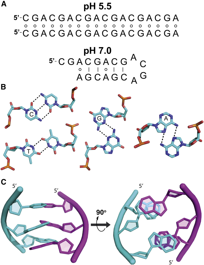
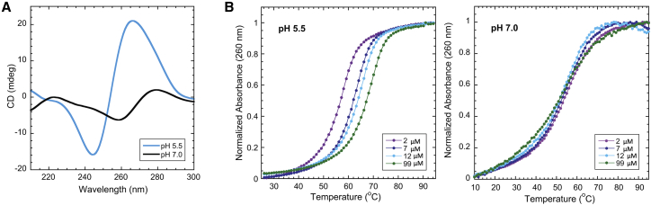
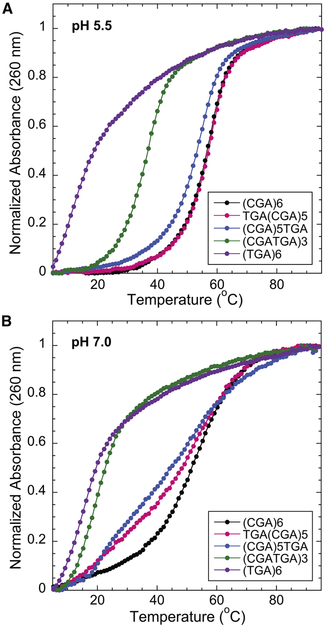
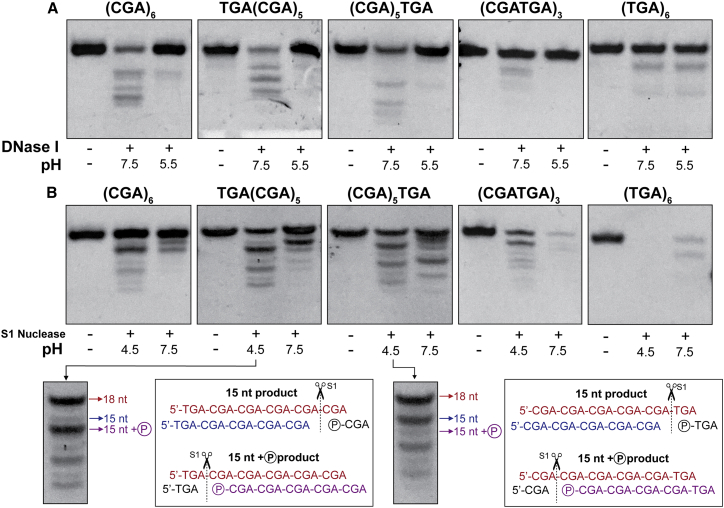
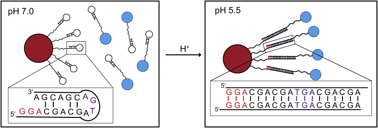
Noncanonical DNA structures that retain programmability and structural predictability are increasingly being used in DNA nanotechnology applications, in which they offer versatility beyond traditional Watson-Crick interactions. The d(CGA) triplet repeat motif is structurally dynamic and can transition between parallel-stranded homo-base paired duplex and antiparallel unimolecular hairpin in a pH-dependent manner. Here, we evaluate the thermodynamic stability and nuclease sensitivity of oligonucleotides composed of the d(CGA) motif and several structurally related sequence variants. These results show that the structural transition resulting from decreasing the pH is accompanied by both a significant energetic stabilization and decreased nuclease sensitivity as unimolecular hairpin structures are converted to parallel-stranded homo-base paired duplexes. Furthermore, the stability of the parallel-stranded duplex form can be altered by changing the 5'-nucleobase of the d(CGA) triplet and the frequency and position of the altered triplets within long stretches of d(CGA) triplets. This work offers insight into the stability and versatility of the d(CGA) triplet repeat motif and provides constraints for using this pH-adaptive structural motif for creating DNA-based nanomaterials.


Figures

Figure 1
Figure 1
d(CGA) triplet motif. (A) (CGA)6 is predicted to form a parallel-stranded homoduplex at pH 5.5 and an antiparallel hairpin at pH 7.0, based on NMR and CD analysis (25, 26, 27, 28). Circles indicate noncanonical base pairing, and solid lines represent canonical base pairing. It is unclear whether the antiparallel hairpin anticipated at pH 7.0 forms with (shown) or without (not shown) A-A homo-base pairs (25). (B) Shown are the following homo-base pairing interactions in the parallel-stranded form of the d(YGA) motif: protonation of cytosine N3 allows formation of a hemiprotonated C-CH+ homo-base pair, T-T homo-base pair, G-G minor groove edge homo-base pair, and A-A Hoogsteen-face homo-base pair. (C) A d(CGA) triplet forming a parallel-stranded homoduplex, shown from the side and also rotated 90° so the 5′ ends go into the page, to show interstrand GA stacking interactions. Schematics in (B) and (C) used Protein Data Bank structures PDB: 4RIM and PDB: 5CV2. To see this figure in color, go online.
Figure 2
Figure 2
(CGA)6 adopts a parallel-stranded duplex or an antiparallel hairpin in response to pH. (A) Shown are CD spectra of 10 μM (CGA)6 at pH 5.5 and pH 7.0. At pH 5.5, the prominent positive band at 265 nm and negative band at 245 nm are consistent with parallel-stranded duplex formation. At pH 7.0, the positive band at 275 nm and negative band at 258 nm are characteristic of antiparallel strands (27). (B) Normalized temperature versus absorbance curves for (CGA)6 show concentration-dependent Tm at pH 5.5 and concentration-independent Tm at pH 7.0, suggesting bi-/multimolecular and largely unimolecular structure formation, respectively. To see this figure in color, go online.
Figure 3
Figure 3
The addition of d(TGA) triplets decreases the Tm of (YGA)6 oligomers. Shown is normalized temperature versus absorbance curves for (YGA)6 oligomers with d(TGA) triplets (n = 6, 3, 1, and 0). (A) At pH 5.5, all variants except (TGA)6 exhibit two-state melting. (B) At pH 7.0, (CGA)6 is the only variant to retain two-state melting. (CGA)5TGA and TGA(CGA)5 have broad melting curves with multiple transitions, whereas (TGA)6 and (CGATGA)3 do not have clear melting transitions, suggesting a lack of stable structure formation. To see this figure in color, go online.
Figure 4
Figure 4
Parallel-stranded duplex enhances nuclease resistance. (A) (YGA)6 variants digested with DNase I at pH 7.5 or 5.5. DNase I preferentially cuts double-stranded DNA, suggesting that the parallel duplexes formed at pH 5.5 are resistant to DNase I degradation. (B) Shown are (YGA)6 variants digested with S1 nuclease at pH 7.5 or 4.5. Red labeled sequences correspond to full length DNA (18-nt), without any nuclease modification. Blue sequences represent the nuclease cleavage product without a terminal phosphate group. The magenta sequence corresponds to the nuclease cleavage product containing a terminal phosphate group, leading to increased mobility. To see this figure in color, go online.
Figure 5
Figure 5
Potential use of (CGA)6 and variants in DNA nanotechnology applications in which pH changes could localize particles together. At pH 7.0, d(CGA) forms hairpin structures, preventing duplex formation, and particles remain separate. At pH 5.5, parallel-stranded duplexes can form and localize particles of interest together. d(TGA) and d(GGA) triplets can be used to ensure desired registration and length of the linking duplex. Linker stretches solely comprised of d(CGA) triplets could be used, but distance between particles of interest could vary. To see this figure in color, go online.

References

    1. Seeman N.C. Nucleic acid junctions and lattices. J. Theor. Biol. 1982;99:237–247. - PubMed
    1. Rothemund P.W.K. Folding DNA to create nanoscale shapes and patterns. Nature. 2006;440:297–302. - PubMed
    1. Jones M.R., Seeman N.C., Mirkin C.A. Nanomaterials. Programmable materials and the nature of the DNA bond. Science. 2015;347:1260901. - PubMed
    1. Seeman N.C. Nanomaterials based on DNA. Annu. Rev. Biochem. 2010;79:65–87. - PMC - PubMed
    1. Wu D., Wang L., Jiang W. DNA nanostructure-based drug delivery nanosystems in cancer therapy. Int. J. Pharm. 2017;533:169–178. - PubMed