. 2020 Nov 12;183(4):968-981.e7.
doi: 10.1016/j.cell.2020.09.016. Epub 2020 Sep 6.

The Immunology of Multisystem Inflammatory Syndrome in Children with COVID-19

Affiliations

The Immunology of Multisystem Inflammatory Syndrome in Children with COVID-19

Camila Rosat Consiglio et al. Cell. .

Abstract

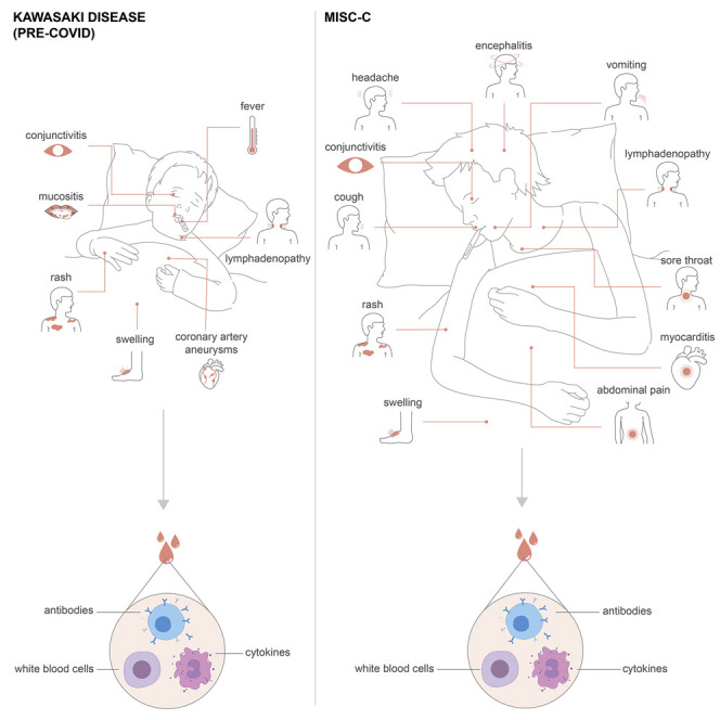
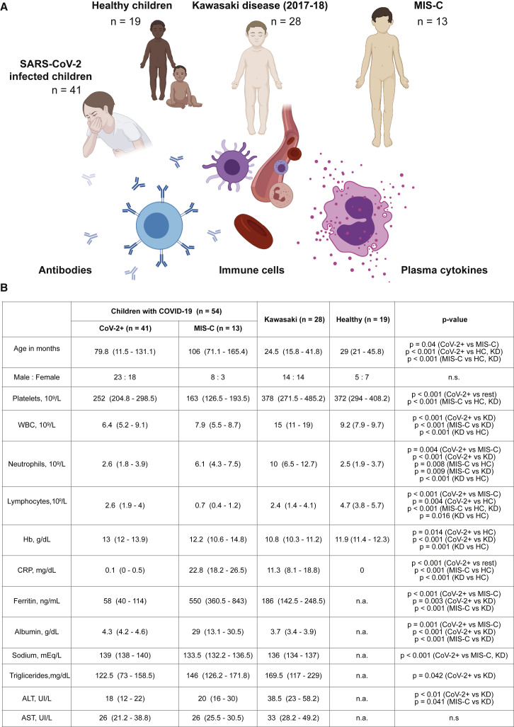
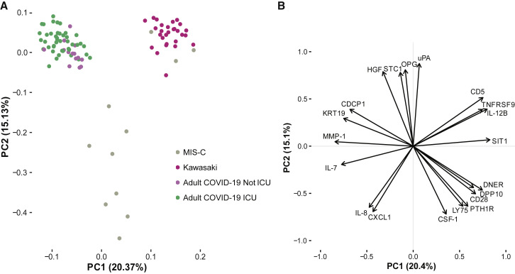
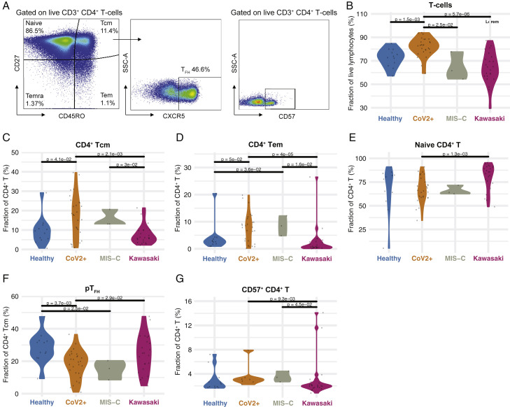
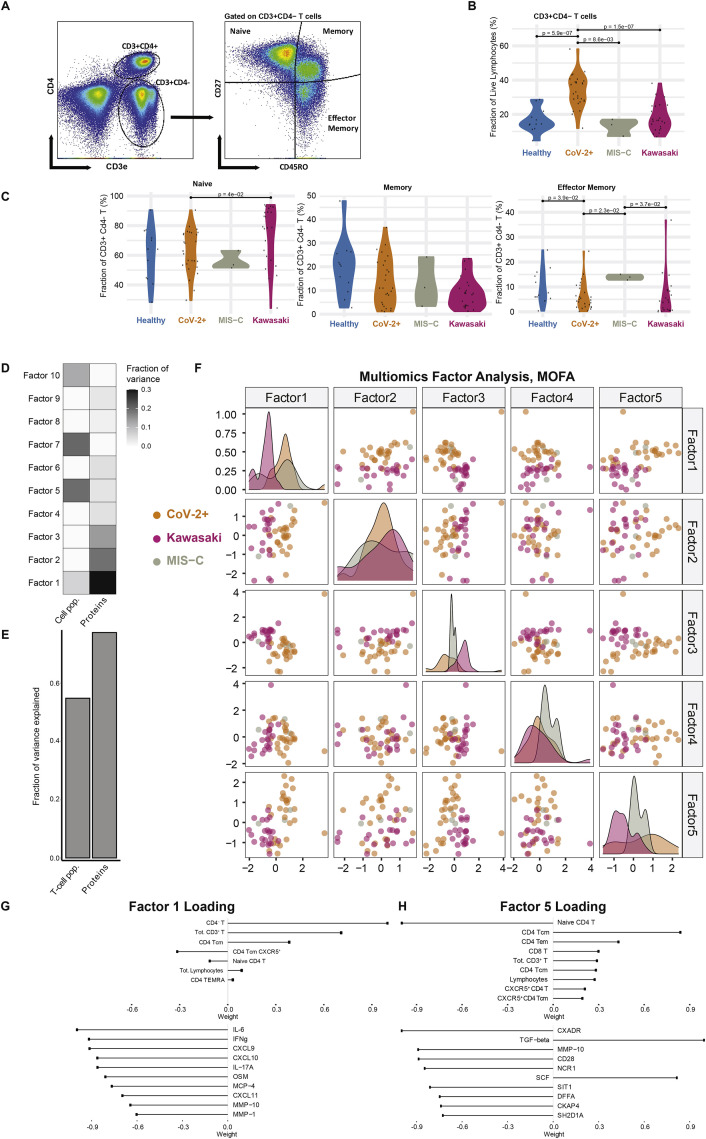
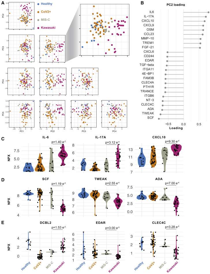
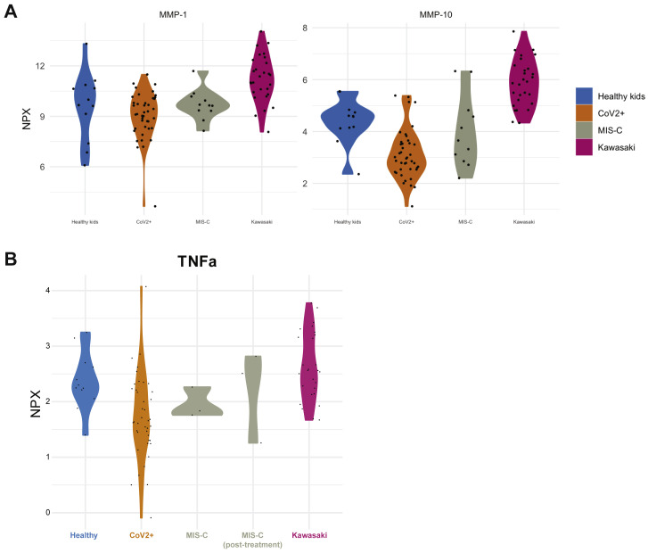
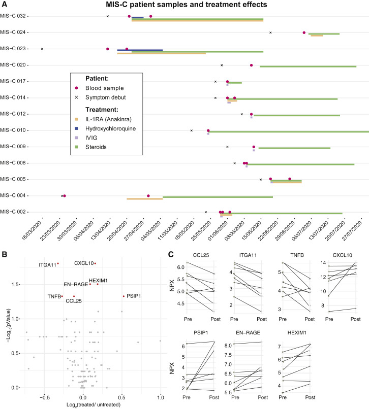
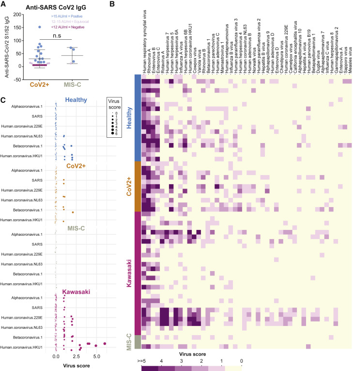
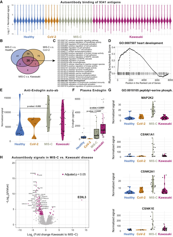
Severe acute respiratory syndrome coronavirus 2 (SARS-CoV-2) infection is typically very mild and often asymptomatic in children. A complication is the rare multisystem inflammatory syndrome in children (MIS-C) associated with COVID-19, presenting 4-6 weeks after infection as high fever, organ dysfunction, and strongly elevated markers of inflammation. The pathogenesis is unclear but has overlapping features with Kawasaki disease suggestive of vasculitis and a likely autoimmune etiology. We apply systems-level analyses of blood immune cells, cytokines, and autoantibodies in healthy children, children with Kawasaki disease enrolled prior to COVID-19, children infected with SARS-CoV-2, and children presenting with MIS-C. We find that the inflammatory response in MIS-C differs from the cytokine storm of severe acute COVID-19, shares several features with Kawasaki disease, but also differs from this condition with respect to T cell subsets, interleukin (IL)-17A, and biomarkers associated with arterial damage. Finally, autoantibody profiling suggests multiple autoantibodies that could be involved in the pathogenesis of MIS-C.

Keywords: COVID-19; IL-17A; Kawasaki disease; MIS-C; SARS-CoV-2; autoantibodies; hyperinflammation in children; multisystem inflammatory syndrome in children; systems immunology.


Conflict of interest statement

Declaration of Interests P.B. and T.L. are founders and shareholders of Cytodelics AB (Stockholm, Sweden). P.B. is an advisor Scailyte AG (Switzerland).

Figures

None
Graphical abstract
Figure 1
Figure 1
Systems-Level Analyses in Children with COVID-19, Kawasaki Disease, and MIS-C (A) Groups of children studied. (B) Clinical parameters shown as median values (25th to 75th centiles). t test (parametric) and Mann-Whitney test (non-parametric) were used to compare mean values across groups of children.
Figure 2
Figure 2
MIS-C Hyperinflammation Differs from Severe Acute COVID-19 Hyperinflammation (A) Principal components 1 and 2 show variation in cytokine profiles among adult COVID-19 patients with severe disease treated in intensive care units (ICU) or not, and children with MIS-C or Kawasaki disease. n = 97 samples included, and 112 unique proteins included in the analysis. (B) Top 20 proteins mostly contributing to the PCs 1–2. See also Table S1.
Figure 3
Figure 3
CD4+ T Cell Subsets in COVID-19 and MIS-C (A) Gating schema to identify CD4+ T cell subsets from peripheral blood mononuclear cells. (B) CD4+ T cells as a fraction of lymphocytes. Black lines indicate statistical tests and p values across indicated populations. (C–G) Fraction of (C) CD4+ Tcm, (D) CD4+ Tem, (E) naive CD4+, (F) pTFH, and (G) CD57+ CD4+ T cell subsets (%) in the indicated patient group. Black lines indicate statistical tests and p values across indicated populations. See also Figure S1.
Figure S1
Figure S1
Multiomics Immune System Differences among Disease Groups, Related to Figure 3 (A) Gating of CD4- T cell subsets. (B) Frequency of CD4- T cells across disease groups. (C) Naive and memory phenotype CD4- T cells. (D) Fraction of variance explained by 10 latent factors, (E) Fraction of variance explained by data type, (F) Samples distributed across the top five latent factors. (G) Top contributing features for factor 1 and (H) factor 5 respectively.
Figure 4
Figure 4
Cytokine Profiles in MIS-C and Kawasaki Disease (A) Principal component analysis of top 5 components explains 58.3% of the variance in concentrations of 133 plasma proteins. PC2 versus 3 separate children with Kawasaki (n = 28) from MIS-C (n = 11), healthy children (n = 12), and SARS-CoV-2+ children without hyperinflammation (n = 41). (B) Proteins explaining (highest loading for) PC2. (C and D) Raw (NPX) values for top (C) positive and (D) negative proteins (loading) in PC2. p values for mean comparisons between MIS-C and Kawasaki disease children. (E) Raw (NPX) values of three proteins significantly different between Kawasaki and MIS-C. p values for mean comparisons between MIS-C and Kawasaki disease children. See also Figure S2 and Table S2.
Figure S2
Figure S2
Related to Figure 4 (A) Plasma MMP-1 and MMP-10 across disease groups. (B) Plasma TNFα levels in patient groups. NPX is Normalized expression level.
Figure 5
Figure 5
Immunomodulatory Treatment in Patients with MIS-C (A) Timelines for individual MIS-C patients (n = 13) indicating date of symptom debut, immunomodulatory treatment regimen, and date of blood sampling. Two patients lacking clinical treatment data were blood sampled prior to treatment. (B) Volcano plot showing fold-change (post-/pre-treatment) versus p value for 7 patients with multiple samples. (C) Top markers of treatment response in MIS-C patients. Fold-change pre/post treatment (p < 0.05, false discovery rate [FDR], 1%). See also Figure S2.
Figure 6
Figure 6
Serological Responses and Prior Immunity in Kawasaki and MIS-C (A) Anti-SARS-CoV-2 S1/S2 IgG antibodies in patients with mild infection (n = 26) and MIS-C (n = 4). (B) VirScan analysis of IgG responses to 93,904 epitopes of 206 viruses in healthy (n = 19), CoV-2 infected (n = 11), Kawasaki disease (n = 27), and children with MIS-C (n = 3). Virus scores is a function of the number of targeted epitopes per virus, and the heatmap shows the top 46 viruses with top average virus scores. (C) The virus scores of coronaviruses available in the VirScan library, shown across individual children divided by disease groups.
Figure 7
Figure 7
Autoantibodies in MIS-C, Kawasaki, and Healthy Children (A) Overall antibody binding intensities against 9,341 antigens from 7,669 human proteins in healthy children (n = 11), CoV-2+ (n = 5), MIS-C (n = 12), and Kawasaki (n = 28) violin plots colored by sample group. (B) Venn diagram showing 26 enriched GO-terms across MIS-C versus healthy, Cov-2+, and Kawasaki disease children. (C) The 26 GO terms enriched in MIS-C versus all other groups listed. (D) GSEA plot for GO:0007507 heart development. (E) Autoantibodies targeting the glycoprotein endoglin (CD105). p value comparing means in healthy children and MIS-C (FDR, 1%). (F) Plasma endoglin levels measured by ELISA in plasma samples. (G) Four candidate antigens bound by autoantibodies across the four patient groups but at highest levels in MIS-C children. (H) Volcano plot showing fold-change differences in autoantibody signals between Kawasaki disease (n = 28) and MIS-C (n = 13). Purple and annotated target antigens have p < 0.05 (FDR, 1%). EDIL3 is the single most overrepresented protein in Kawasaki disease. See also Table S3.

Comment in

  • The Mystery of MIS-C Post-SARS-CoV-2 Infection.
    Brodsky NN, Ramaswamy A, Lucas CL. Brodsky NN, et al. Trends Microbiol. 2020 Dec;28(12):956-958. doi: 10.1016/j.tim.2020.10.004. Epub 2020 Oct 14. Trends Microbiol. 2020. PMID: 33190685 Free PMC article.

References

    1. Anft M., Paniskaki K., Blazquez-Navarro A., Doevelaar A.A.N., Seibert F., Hoelzer B., Skrzypczyk S., Kohut E., Kurek J., Zapka J., et al. COVID-19 progression is potentially driven by T cell immunopathogenesis. medRxiv. 2020 doi: 10.1101/2020.04.28.20083089. - DOI
    1. Argelaguet R., Arnol D., Bredikhin D., Deloro Y., Velten B., Marioni J.C., Stegle O. MOFA+: a probabilistic framework for comprehensive integration of structured single-cell data. bioRxiv. 2019 doi: 10.1101/837104. - DOI - PMC - PubMed
    1. Bouhaddou M., Memon D., Meyer B., White K.M., Rezelj V.V., Marrero M.C., Polacco B.J., Melnyk J.E., Ulferts S., Kaake R.M., et al. The Global Phosphorylation Landscape of SARS-CoV-2 Infection. Cell. 2020;182:685–712. - PMC - PubMed
    1. Brodin P. Why is COVID-19 so mild in children? Acta Paediatr. 2020;109:1082–1083. - PubMed
    1. Cheung E.W., Zachariah P., Gorelik M., Boneparth A., Kernie S.G., Orange J.S., Milner J.D. Multisystem Inflammatory Syndrome Related to COVID-19 in Previously Healthy Children and Adolescents in New York City. JAMA. 2020;324:294–296. - PMC - PubMed

Publication types

MeSH terms

Supplementary concepts