Review
. 2020 Sep 21;9(9):2132.
doi: 10.3390/cells9092132.

Microglia and Macrophages in the Pathological Central and Peripheral Nervous Systems

Affiliations
Review

Microglia and Macrophages in the Pathological Central and Peripheral Nervous Systems

Naoki Abe et al. Cells. .

Abstract

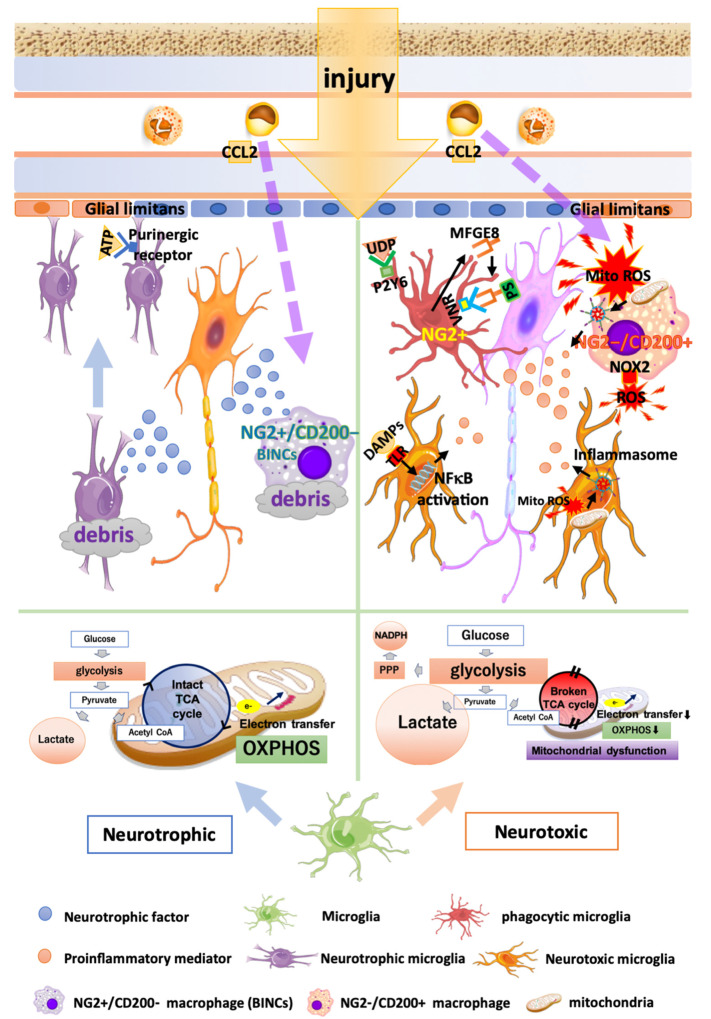
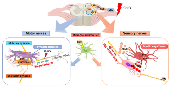
Microglia, the immunocompetent cells in the central nervous system (CNS), have long been studied as pathologically deteriorating players in various CNS diseases. However, microglia exert ameliorating neuroprotective effects, which prompted us to reconsider their roles in CNS and peripheral nervous system (PNS) pathophysiology. Moreover, recent findings showed that microglia play critical roles even in the healthy CNS. The microglial functions that normally contribute to the maintenance of homeostasis in the CNS are modified by other cells, such as astrocytes and infiltrated myeloid cells; thus, the microglial actions on neurons are extremely complex. For a deeper understanding of the pathophysiology of various diseases, including those of the PNS, it is important to understand microglial functioning. In this review, we discuss both the favorable and unfavorable roles of microglia in neuronal survival in various CNS and PNS disorders. We also discuss the roles of blood-borne macrophages in the pathogenesis of CNS and PNS injuries because they cooperatively modify the pathological processes of resident microglia. Finally, metabolic changes in glycolysis and oxidative phosphorylation, with special reference to the pro-/anti-inflammatory activation of microglia, are intensively addressed, because they are profoundly correlated with the generation of reactive oxygen species and changes in pro-/anti-inflammatory phenotypes.

Keywords: NG2; brain infarction; carbon monoxide poisoning; macrophage; peripheral nerve injury; traumatic brain injury.


Conflict of interest statement

The authors declare no conflict of interest.

Figures

Figure 1
Figure 1
Multiple functions of microglia and macrophages in brain injury. Neurotrophic microglia and macrophages (left half) exhibit an intact TCA cycle and stable mitochondrial OXPHOS. Microglia that have migrated to the injury site release anti-inflammatory cytokines and neurotrophic factors, thus encouraging wound healing and debris clearance. Neuroprotective infiltrated macrophages called BINCs (brain Iba1+/NG2+ cells) express a variety of neuroprotective factors. Neurotoxic microglia and macrophages (right half) produce energy in a glycolysis-dependent manner and exhibit increased lactate production, glucose uptake, and pentose phosphate pathway (PPP). DAMPs recognized by TLR stimulate NFκB pathways, leading to an increased expression of proinflammatory mediators. Microglia phagocytose viable neurons by recognizing opsonized PS via VNRs and the humoral “eat-me” signal UDP, through P2Y6. Phagocytic microglia and macrophages express the phagocytosis marker CD68 and NG2. Neurotoxic infiltrated macrophages (NG2/CD200+ macrophages) release a greater amount of MitoROS, IL1β, and NOX2 compared with microglia. MitoROS not only directly damages the brain tissues, but also induces proinflammatory reactions by inducing the formation of inflammasomes.
Figure 2
Figure 2
Functions of spinal microglia in peripheral nerve injury. When the peripheral nerve is injured, the colony-stimulating factor 1 (CSF1) is rapidly induced in DRG neurons. CSF1 transported to the spinal dorsal horn acts on the CSF1 receptor (CSF1R) of microglia, for their proliferation and activation. In the dorsal horn, the interferon regulatory factor 5 (IRF5) and IRF8, which are transcription factors, are induced in the activated microglia, followed by the release of several cytokines (including the tumor necrosis factors α, interleukin 1β, and the brain-derived neurotrophic factor), via which hypersensitivity is induced and maintained. Microglia activated by CSF1 in the ventral horn block signals from inhibitory synapses by synaptic stripping and induce the expression of several factors, which stimulates the degeneration and regeneration of injured nerves.
Figure 3
Figure 3
Effects of BU on cell metabolism. The effects of BU on cell metabolism were investigated in primary microglial cells and macrophages using the Seahorse Mito-Stress Test, which assesses OCR and ECAR. In the Mito-Stress Test, to evaluate mitochondrial respiration and glycolysis, various mitochondrial function inhibitors (1, Oligomycin; 2, FCCP; 3, Rotenon and antimycin) are automatically and sequentially added to the cells. BU inhibited mitochondrial ATP production (OXPHOS) but did not affect mitochondrial membrane permeability or coupling efficacy (ATP production divided by basal respiration). BU also prevented the compensatory enhancement of glycolytic activity after the inhibition of mitochondrial ATP synthase (Oligomycin). Based on these results, BU seems to inhibit both OXPHOS and glycolytic pathways without causing mitochondrial dysfunction.

References

    1. Ginhoux F., Greter M., Leboeuf M., Nandi S., See P., Gokhan S., Mehler M.F., Conway S.J., Ng L.G., Stanley E.R., et al. Fate mapping analysis reveals that adult microglia derive from primitive macrophages. Science. 2010;330:841–845. doi: 10.1126/science.1194637. - DOI - PMC - PubMed
    1. Kierdorf K., Erny D., Goldmann T., Sander V., Schulz C., Perdiguero E.G., Wieghofer P., Heinrich A., Riemke P., Holscher C., et al. Microglia emerge from erythromyeloid precursors via Pu.1- and Irf8-dependent pathways. Nat. Neurosci. 2013;16:273–280. doi: 10.1038/nn.3318. - DOI - PubMed
    1. Dudvarski Stankovic N., Teodorczyk M., Ploen R., Zipp F., Schmidt M.H.H. Microglia-blood vessel interactions: A double-edged sword in brain pathologies. Acta Neuropathol. 2016;131:347–363. doi: 10.1007/s00401-015-1524-y. - DOI - PubMed
    1. Kettenmann H., Kirchhoff F., Verkhratsky A. Microglia: New roles for the synaptic stripper. Neuron. 2013;77:10–18. doi: 10.1016/j.neuron.2012.12.023. - DOI - PubMed
    1. Inoue K., Tsuda M. Microglia in neuropathic pain: Cellular and molecular mechanisms and therapeutic potential. Nat. Rev. Neurosci. 2018;19:138–152. doi: 10.1038/nrn.2018.2. - DOI - PubMed

MeSH terms

Substances

LinkOut - more resources