

Non-Invasive Analysis of Actinic Keratosis before and after Topical Treatment Using a Cold Stimulation and Near-Infrared Spectroscopy
- PMID: 32967260
- PMCID: PMC7560046
- DOI: 10.3390/medicina56090482
Non-Invasive Analysis of Actinic Keratosis before and after Topical Treatment Using a Cold Stimulation and Near-Infrared Spectroscopy
Abstract
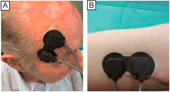
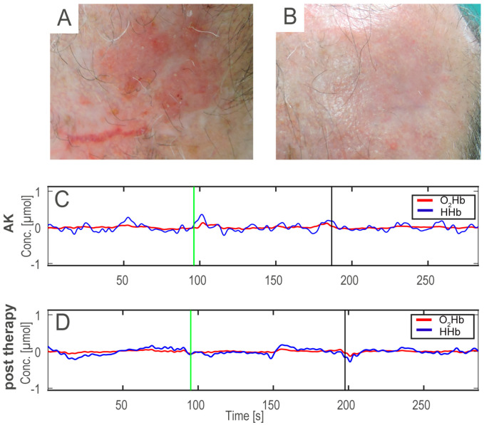
Background and objectives: The possible evolution of actinic keratoses (AKs) into invasive squamous cell carcinomas (SCC) makes their treatment and monitoring essential. AKs are typically monitored before and after treatment only through a visual analysis, lacking a quantitative measure to determine treatment effectiveness. Near-infrared spectroscopy (NIRS) is a non-invasive measure of the relative change of oxy-hemoglobin and deoxy-hemoglobin (O2Hb and HHb) in tissues. The aim of our study is to determine if a time and frequency analysis of the NIRS signals acquired from the skin lesion before and after a topical treatment can highlight quantitative differences between the AK skin lesion area. Materials and Methods: The NIRS signals were acquired from the skin lesions of twenty-two patients, with the same acquisition protocol: baseline signals, application of an ice pack near the lesion, removal of ice pack and acquisition of vascular recovery. We calculated 18 features from the NIRS signals, and we applied multivariate analysis of variance (MANOVA) to compare differences between the NIRS signals acquired before and after the therapy. Results: The MANOVA showed that the features computed on the NIRS signals before and after treatment could be considered as two statistically separate groups, after the ice pack removal. Conclusions: Overall, the NIRS technique with the cold stimulation may be useful to support non-invasive and quantitative lesion analysis and regression after a treatment. The results provide a baseline from which to further study skin lesions and the effects of various treatments.
Keywords: actinic keratosis; field cancerization; near-infrared spectroscopy; signal processing.
Conflict of interest statement
The authors declare no conflict of interest.
Figures

Similar articles
-
Non-invasive analysis of actinic keratosis using a cold stimulation and near-infrared spectroscopy.Annu Int Conf IEEE Eng Med Biol Soc. 2019 Jul;2019:467-470. doi: 10.1109/EMBC.2019.8857279. Annu Int Conf IEEE Eng Med Biol Soc. 2019. PMID: 31945939
-
Non-invasive monitoring of subclinical and clinical actinic keratosis of face and scalp under topical treatment with ingenol mebutate gel 150 mcg/g by means of reflectance confocal microscopy and optical coherence tomography: New perspectives and comparison of diagnostic techniques.J Biophotonics. 2019 Jul;12(7):e201800391. doi: 10.1002/jbio.201800391. Epub 2019 Mar 20. J Biophotonics. 2019. PMID: 30653833
-
Using computational learning for non-melanoma skin cancer and actinic keratosis near-infrared hyperspectral signature classification.Photodiagnosis Photodyn Ther. 2024 Oct;49:104269. doi: 10.1016/j.pdpdt.2024.104269. Epub 2024 Jul 11. Photodiagnosis Photodyn Ther. 2024. PMID: 39002835
-
Prevalence, Discontinuation Rate, and Risk Factors for Severe Local Site Reactions with Topical Field Treatment Options for Actinic Keratosis of the Face and Scalp.Medicina (Kaunas). 2019 Apr 4;55(4):92. doi: 10.3390/medicina55040092. Medicina (Kaunas). 2019. PMID: 30987411 Free PMC article. Review.
-
Actinic keratosis and squamous cell carcinoma: clinical and pathological features.G Ital Dermatol Venereol. 2015 Aug;150(4):379-84. Epub 2015 Jun 23. G Ital Dermatol Venereol. 2015. PMID: 26099352 Review.
Cited by
-
AKASI and Near-Infrared Spectroscopy in the combined effectiveness evaluation of an actinic keratoses preventive product in immunocompetent and immunocompromised patients.Front Med (Lausanne). 2022 Sep 7;9:987696. doi: 10.3389/fmed.2022.987696. eCollection 2022. Front Med (Lausanne). 2022. PMID: 36160127 Free PMC article.
References
-
- Fargnoli M.C., Altomare G., Benati E., Borgia F., Broganelli P., Carbone A., Chimenti S., Donato S., Girolomoni G., Micali G., et al. Prevalence and risk factors of actinic keratosis in patients attending Italian dermatology clinics. Eur. J. Dermatol. 2017;27:599–608. doi: 10.1684/ejd.2017.3126. - DOI - PubMed
MeSH terms
Substances
LinkOut - more resources
Full Text Sources
Medical
Research Materials
