. 2020 Sep 21;21(18):6935.
doi: 10.3390/ijms21186935.

Sappanone A Prevents Left Ventricular Dysfunction in a Rat Myocardial Ischemia Reperfusion Injury Model

Affiliations

Sappanone A Prevents Left Ventricular Dysfunction in a Rat Myocardial Ischemia Reperfusion Injury Model

Woori Jo et al. Int J Mol Sci. .

Abstract

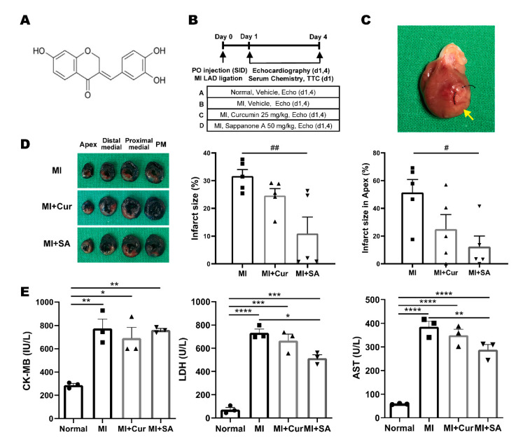
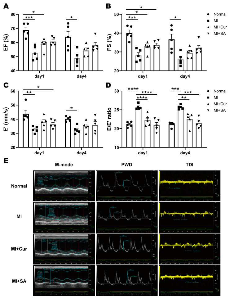
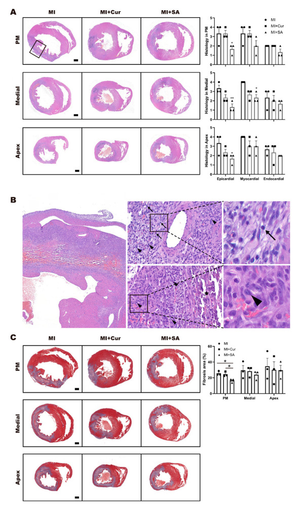
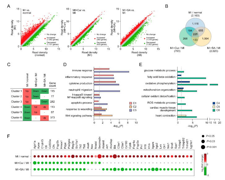
The incidence of myocardial infarction, among the causes of cardiovascular morbidity and mortality, is increasing globally. In this study, left ventricular (LV) dysfunction, including LV systolic and diastolic function, was investigated in a rat myocardial ischemia/reperfusion injury model with echocardiography. The homoisoflavanone sappanone A is known for its anti-inflammatory effects. Using echocardiography, we found that sappanone A administration significantly improved LV systolic and diastolic function in a rat myocardial ischemia/reperfusion injury model, especially in the early phase development of myocardial infarction. Based on myocardial infarct size, serum cardiac marker assay, and histopathological evaluation, sappanone A showed higher efficacy at the doses used in our experiments than curcumin and was evaluated for its potential to improve LV function.

Keywords: acute myocardial infarction; coronary artery ligation; diastolic function; myocardial ischemia/reperfusion injury; sappanone A.


Conflict of interest statement

The authors have declared no conflict of interest.

Figures

Figure 1
Figure 1
The experimental design and the positive effect on the infarct size and serum cardiac marker. (A) Chemical structure of sappanone A. (B) The animals were divided into four groups: Group A, Normal control; Group B, MI+Vehicle; Group C, MI+Curcumin 25 mg/kg; Group D, MI+Sappanone A 50 mg/kg. (C) Representative gross photograph of a heart on day 1 after MI surgery and the infarct area (yellow arrow). (D) Representative photographs of left ventricle slices of the groups and the infarct size of the left ventricles of the total and apex areas in each group (n = 5/group, right). # p < 0.05 and ## p < 0.01 by one-way ANOVA test with Tukey’s post hoc correction. (E) Serum chemistry results of creatine kinase MB isozyme (CK-MB, n = 3/group), lactate dehydrogenase (LDH, n = 3/group), and aspartate aminotransferase (AST, n = 3/group) on day 1 after MI surgery. ** p < 0.01, *** p < 0.001, **** p < 0.0001 by two-way ANOVA test with Tukey’s post hoc correction.
Figure 2
Figure 2
Echocardiographic results (n = 5/group) and the representative echocardiographic images of rat left ventricle on day 4 after MI surgery. Sappanone A significantly improved left ventricular (LV) systolic and diastolic function. (A) Ejection fraction, EF; (B) Fractional shortening, FS; (C) Early relaxation velocity on tissue Doppler, E′; (D) E/E′ ratio of LV diastolic function; (E) Representative echocardiographic imaging on day 4 after MI surgery. * p < 0.05, ** p < 0.01, *** p < 0.001, **** p < 0.0001 by two-way ANOVA tests with Tukey’s post hoc correction.
Figure 3
Figure 3
Histopathological images and results in each left ventricle area on day 4 after MI surgery. Representative photomicrographs are shown in hematoxylin and eosin and incidence of histopathological changes (A, ×12.5) and representative H&E photomicrographs with high magnification (B, ×40, ×400, ×1500). Mature lymphocyte (arrows) and mononuclear cells (arrow heads) are shown in myocardial region (Top, ×400) and mononuclear cells (arrow heads) with purulent (asterisk) are shown in endocardial region (Bottom, ×400). Representative cell image of lymphocyte (arrow) and mononuclear cell (arrow head) can be identified with high magnification (×1500). Representative photomicrographs in Masson’s trichrome and fibrosis area percentage (C, ×12.5). * p < 0.05, indicate statistically significant differences by one-way ANOVA with Tukey’s post hoc correction. Grading of histopathological changes in each tissue area (papillary muscle, medial, and apex) of the rat left ventricle. Grades 1, 2, 3, and 4 show minimal, slight, moderate, and severe pathological changes, respectively. Values are mean ± standard error of the mean (SEM, n = 3).
Figure 4
Figure 4
Cellular processes affected by sappanone A. (A) Scatter plots of three comparisons (MI versus normal, MI + Curcumin versus MI, and MI + Sappanone A versus MI). (B) Relationships among differentially expressed genes (DEGs). (C) Clusters (C1-6) of the genes affected by curcumin or sappanone A. Red and green denote up- and down-regulation, respectively. The number of DEGs in each cluster is denoted. (D,E) Cellular processes represented by DEGs in C1-6. X-axis, −log10(P) where P is the enrichment p-value calculated in DAVID software. (F) DEGs involved in inflammatory responses.
Figure 5
Figure 5
Inhibitory effects of Sappanone A administration on the MI-related processes. (A) Network model describing interactions among signaling pathways. Arrows, activation in signaling. “+p”, phosphorylation. (B) Confirmation of the predominant downregulation of the eight representative genes involved in the inflammatory responses and apoptosis by RT-PCR. The expression levels were normalized with respect to those in the control group. The normalized data are expressed as the mean ± SEM (n = 4 per group). * p < 0.05, ** p < 0.01, *** p < 0.001, **** p < 0.0001 by two-way ANOVA tests with Tukey’s post hoc correction.

References

    1. Jayaraj J.C., Davatyan K., Subramanian S.S., Priya J. Epidemiology of myocardial infarction. In: Pamukçu B., editor. Myocardial Infarction. IntechOpen; London, UK: 2018.
    1. Mor-Avi V., Lang R.M., Badano L.P., Belohlavek M., Cardim N.M., Derumeaux G., Galderisi M., Marwick T., Nagueh S.F., Sengupta P.P., et al. Current and evolving echocardiographic techniques for the quantitative evaluation of cardiac mechanics: ASE/EAE consensus statement on methodology and indications endorsed by the Japanese Society of Echocardiography. Eur. J. Echocardiogr. 2011;12:167–205. doi: 10.1093/ejechocard/jer021. - DOI - PubMed
    1. Lee K.S., Marwick T.H., Cook S.A., Go R.T., Fix J.S., James K.B., Sapp S.K., MacIntyre W.J., Thomas J.D. Prognosis of patients with left ventricular dysfunction, with and without viable myocardium after myocardial infarction. Relative efficacy of medical therapy and revascularization. Circulation. 1994;90:2687–2694. doi: 10.1161/01.CIR.90.6.2687. - DOI - PubMed
    1. Sanders D., Dudley M., Groban L. Diastolic dysfunction, cardiovascular aging, and the anesthesiologist. Anesthesiol. Clin. 2009;27:497–517. doi: 10.1016/j.anclin.2009.07.008. - DOI - PMC - PubMed
    1. Moller J.E., Pellikka P.A., Hillis G.S., Oh J.K. Prognostic importance of diastolic function and filling pressure in patients with acute myocardial infarction. Circulation. 2006;114:438–444. doi: 10.1161/CIRCULATIONAHA.105.601005. - DOI - PubMed

MeSH terms

LinkOut - more resources