

Adding exogenous biglycan or decorin improves tendon formation for equine peritenon and tendon proper cells in vitro
- PMID: 32967653
- PMCID: PMC7513506
- DOI: 10.1186/s12891-020-03650-2
Adding exogenous biglycan or decorin improves tendon formation for equine peritenon and tendon proper cells in vitro
Abstract
Background: Tendon injuries amount to one of the leading causes of career-ending injuries in horses due to the inability for tendon to completely repair and the high reinjury potential. As a result, novel therapeutics are necessary to improve repair with the goal of decreasing leg lameness and potential reinjury. Small leucine-rich repeat proteoglycans (SLRPs), a class of regulatory molecules responsible for collagen organization and maturation, may be one such therapeutic to improve tendon repair. Before SLRP supplementation can occur in vivo, proper evaluation of the effect of these molecules in vitro needs to be assessed. The objective of this study was to evaluate the effectiveness of purified bovine biglycan or decorin on tendon proper and peritenon cell populations in three-dimensional tendon constructs.
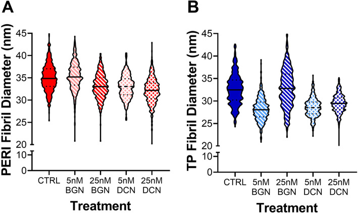
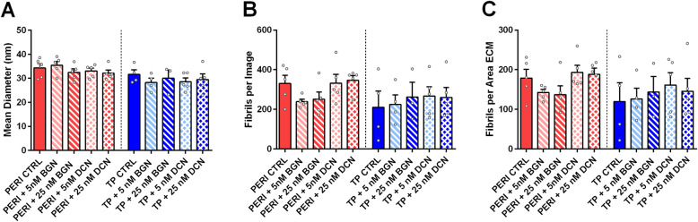
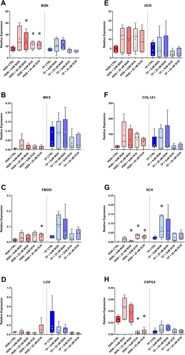
Methods: Equine tendon proper or peritenon cell seeded fibrin three-dimensional constructs were supplemented with biglycan or decorin at two concentrations (5 nM or 25 nM). The functionality and ultrastructural morphology of the constructs were assessed using biomechanics, collagen content analysis, transmission electron microscopy (TEM), and gene expression by real time - quantitative polymerase chain reaction (RT-qPCR).
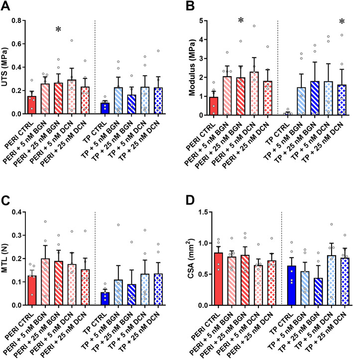
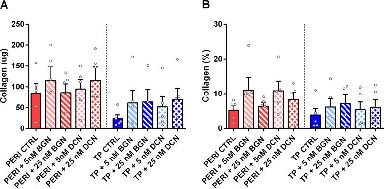
Results: SLRP supplementation affected both tendon proper and peritenon cells-seeded constructs. With additional SLRPs, material and tensile properties of constructs strengthened, though ultrastructural analyses indicated production of similar-sized or smaller fibrils. Overall expression of tendon markers was bolstered more in peritenon cells supplemented with either SLRP, while supplementation of SLRPs to TP cell-derived constructs demonstrated fewer changes in tendon and extracellular matrix markers. Moreover, relative to non-supplemented tendon proper cell-seeded constructs, SLRP supplementation of the peritenon cells showed increases in mechanical strength, material properties, and collagen content.
Conclusions: The SLRP-supplemented peritenon cells produced constructs with greater mechanical and material properties than tendon proper seeded constructs, as well as increased expression of matrix assembly molecules. These findings provide evidence that SLRPs should be further investigated for their potential to improve tendon formation in engineered grafts or post-injury.
Keywords: Biglycan; Decorin; Equine; Peritenon; Tendon; Three-dimensional construct.
Conflict of interest statement
The authors declare that they have no competing interests.
Figures
References
-
- Tipton TE, Ray CS, Hand DR. Superficial digital flexor tendonitis in cutting horses: 19 cases (2007-2011) J Am Vet Med A. 2013;243:1162–1165. - PubMed
-
- Thorpe CT, Glegg PD, Birch HL. A review of tendon injury: why is the equine superficial digital flexor tendon most at risk? Equine Vet J. 2010;42(2):174–180. - PubMed
-
- Halper J. Connective tissue disorders in domestic animals. Adv Exp Med Biol. 2014;802:231–240. - PubMed
-
- Mienaltowski MJ, Birk DE. Structure, physiology, and biochemistry of collagens. Adv Exp Med Biol. 2014;802:5–29. - PubMed
MeSH terms
Substances
LinkOut - more resources
Full Text Sources
Research Materials
