Comparative Study
. 2020 Sep 23;15(1):436.
doi: 10.1186/s13018-020-01935-9.

Evaluation of three-dimensional acromiohumeral distance in the standing position and comparison with its conventional measuring methods

Affiliations
Comparative Study

Evaluation of three-dimensional acromiohumeral distance in the standing position and comparison with its conventional measuring methods

Yuki Yoshida et al. J Orthop Surg Res. .

Abstract

Background: Narrowing of the acromiohumeral distance (AHD) implies a rotator cuff tear. However, conventional AHD measurements using two-dimensional (2D) imaging or with the patient in the supine position might differ from that while standing during daily activity. This study aimed to evaluate the three-dimensional (3D) actual distance between the acromion and humeral head in the standing position and compare the AHD values with those obtained using conventional measuring methods.

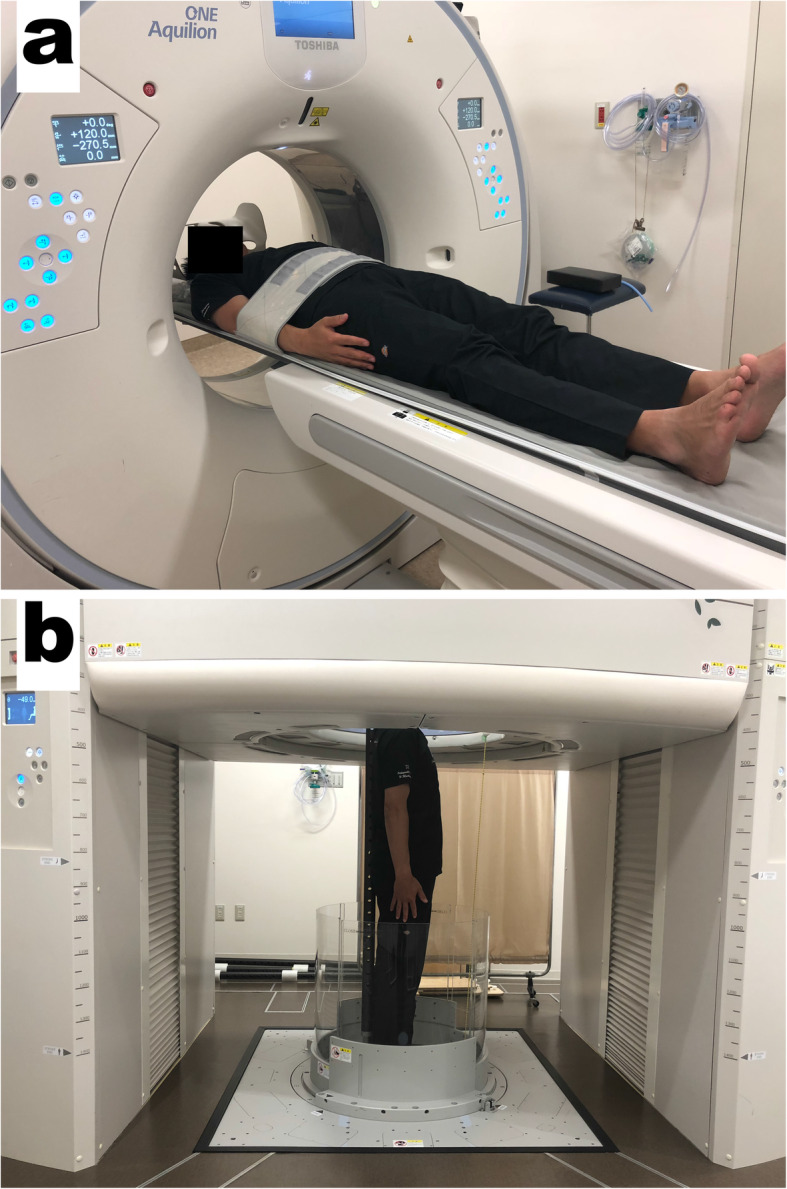
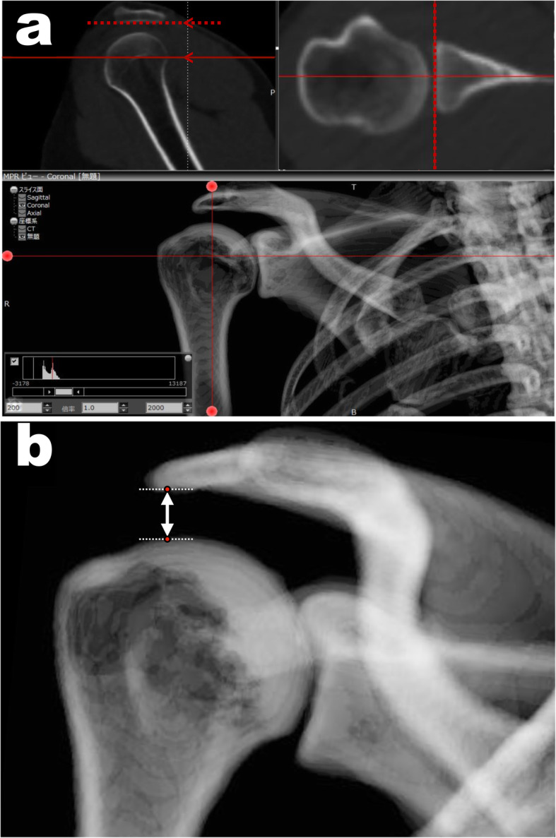
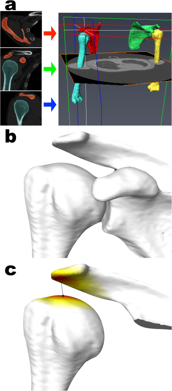
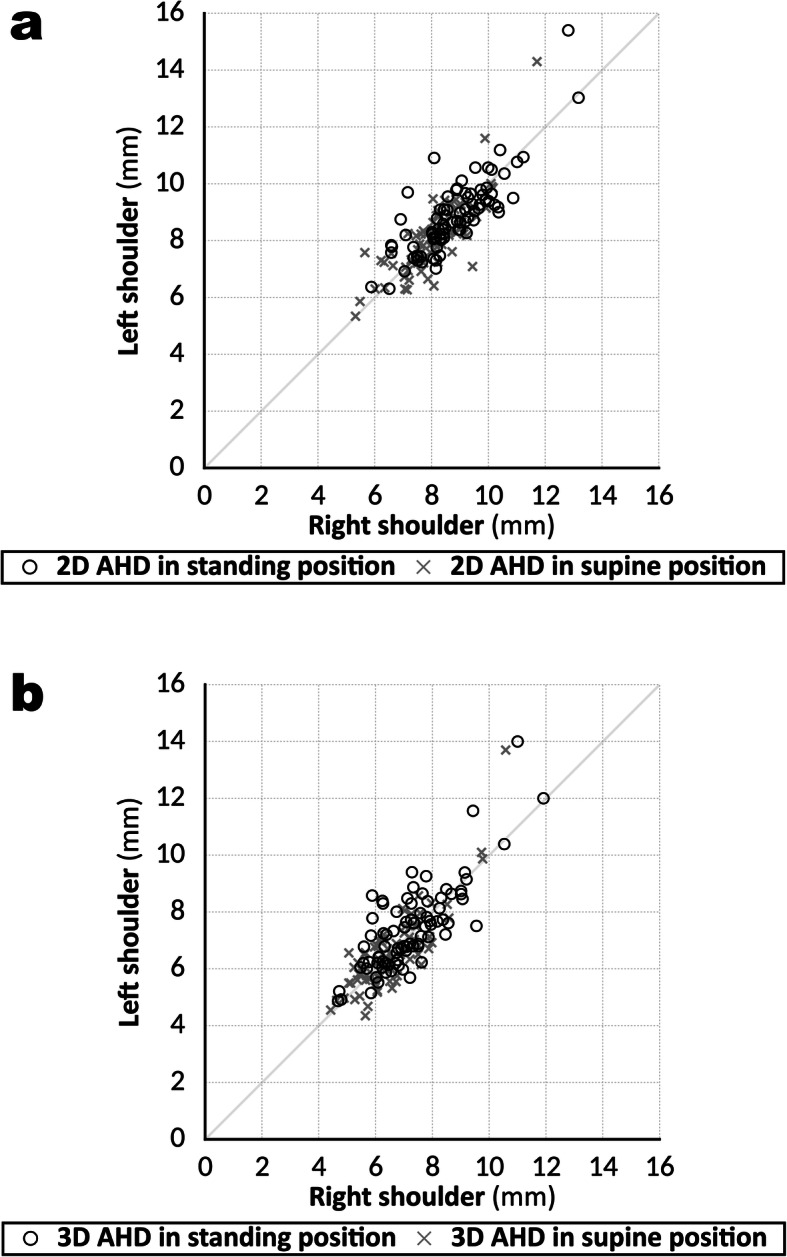
Methods: Computed tomography (CT) images of 166 shoulders from 83 healthy volunteers (31 male and 52 female; mean age 40.1 ± 5.8 years; age range, 30-49 years) were prospectively acquired in the supine and standing positions using conventional and upright CT scanners, respectively. The minimum distance between the acromion and humeral head on the 3D surface models was considered as the 3D AHD. We measured the 2D AHD on anteroposterior digitally reconstructed radiographs. The AHD values were compared between the supine and standing positions and between the 2D and 3D measurements.

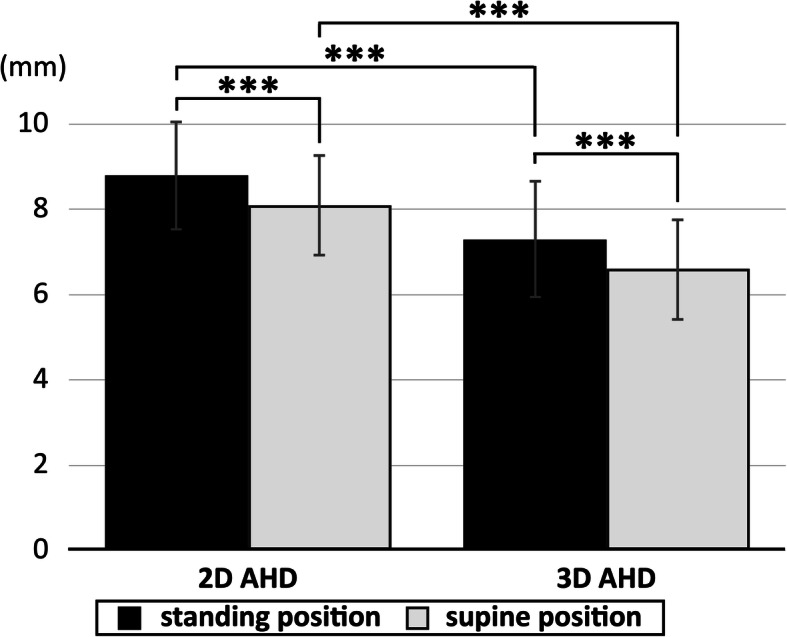
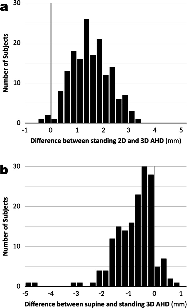
Results: The mean values of 2D AHD were 8.8 ± 1.3 mm (range, 5.9-15.4 mm) in the standing position and 8.1 ± 1.2 mm (range, 5.3-14.3 mm) in the supine position. The mean values of 3D AHD were 7.3 ± 1.4 mm (range, 4.7-14.0 mm) in the standing position and 6.6 ± 1.2 mm (range, 4.4-13.7 mm) in the supine position. The values of 3D AHD were significantly lower than those of 2D AHDs in both the standing and supine positions (P < 0.001). The values of 2D and 3D AHDs were significantly lower in the supine position than in the standing position (P < 0.001).

Conclusions: This study evaluated the 3D AHD of normal shoulders in the standing position using an upright CT scanner. The present results indicated that assessments in the supine position can underestimate the value of the AHD compared with those made in the standing position and that assessments using 2D analysis can overestimate the value.

Keywords: Acromiohumeral distance; Acromiohumeral interval; Digitally reconstructed radiographs; Normal shoulder; Position; Upright computed tomography.


Conflict of interest statement

Masahiro Jinzaki has received a grant from Canon Medical Systems. However, Canon Medical Systems was not involved in the design and conduct of the study; in the collection, analysis, and interpretation of the data; or in the preparation, review, and approval of the manuscript. The remaining authors have no conflicts of interest to declare.

Figures

Fig. 1
Fig. 1
320-row conventional and upright computed tomography (CT) scanners. CT images of the bilateral shoulders were obtained with the shoulders adducted and the arms held in a neutral position, both in the supine position using a conventional scanner (a) and in the standing position using an upright scanner (b)
Fig. 2
Fig. 2
The process of measuring the two-dimensional acromiohumeral distances (2D AHD). a Creating true anteroposterior shoulder digitally reconstructed radiograph (DRR) image from the CT data using ZedView software. Scapula rotation was corrected to align parallel to the inferior aspect of the acromion in sagittal view and align parallel to the face of the glenoid in axial view. b The acromiohumeral distance (AHD) was measured using a two-dimensional (2D) approach on DRR images of the anteroposterior shoulder reconstructed from the CT scans. The 2D AHD was defined as the shortest distance from the dense cortical bone at the inferior aspect of the acromion to the most proximal articular cortex of the humeral head (white line with arrow)
Fig. 3
Fig. 3
The process of calculating the three-dimensional acromiohumeral distances (3D AHD). a Creating 3D surface models of the scapula and humerus from the CT data to observe three views of multiplanar reformatting carefully using AVIZO software. b Bone surface model of the scapula and proximal humerus. c After removing the glenoid and coracoid parts of the surface model, the 3D AHD was automatically measured as the minimum distance between the acromion and humeral head on the software. The red areas indicate where the distance between the acromion and the humeral head is at a minimum
Fig. 4
Fig. 4
Differences in acromiohumeral distances (AHD) between 2D and 3D measurements and between the supine and standing positions. The 3D AHD values were significantly lower than the 2D AHD values in both the standing and supine positions. The 2D and 3D AHD values were significantly lower in the supine position than in the standing position. ***P < 0.001
Fig. 5
Fig. 5
Histogram of the individual differences in acromiohumeral distance (AHD). a Histogram of the individual differences in AHD in the standing position between the 2D and 3D measurements. Positive values indicate the 2D value is greater than the 3D value. b Histogram of the individual differences in 3D AHD between the supine and standing positions. Positive values indicate that the standing value is greater than the supine value. The differences varied widely
Fig. 6
Fig. 6
Linear regression plots of acromiohumeral distance (AHD) values compared between the right and left shoulders. a Linear regression plots of acromiohumeral distance (AHD) values measured in two dimensions in the supine and standing positions, compared between the right and left shoulders. The values showed a strong correlation between the sides (standing: R = 0.794, P < 0.001; supine: R = 0.780, P < 0.001). b Linear regression plots for AHD values measured in three dimensions in the supine and standing positions, compared between the right and left shoulders. The values showed a strong correlation between the sides (standing: R = 0.711, P < 0.001; supine: R = 0.742, P < 0.001)

Similar articles

Cited by

References

    1. Cotton RE, Rideout DF. Tears of the humeral rotator cuff; a radiological and pathological necropsy survey. J Bone Joint Surg Br. 1964;46:314–328. doi: 10.1302/0301-620X.46B2.314. - DOI - PubMed
    1. Golding FC. The shoulder--the forgotten joint. Br J Radiol. 1962;35:149–158. doi: 10.1259/0007-1285-35-411-149. - DOI - PubMed
    1. Petersson CJ, Redlund-Johnell I. The subacromial space in normal shoulder radiographs. Acta Orthop Scand. 1984;55:5758. doi: 10.3109/17453678408992312. - DOI - PubMed
    1. Weiner DS, Macnab I. Superior migration of the humeral head. A radiological aid in the diagnosis of tears of the rotator cuff. J Bone Joint Surg Br. 1970;52:524–527. doi: 10.1302/0301-620X.52B3.524. - DOI - PubMed
    1. Bernhardt GA, Glehr M, Zacherl M, Wurnig C, Gruber G. Observer variability in the assessment of the acromiohumeral interval using anteroposterior shoulder radiographs. Eur J Orthop Surg Traumatol. 2013;23:185–190. doi: 10.1007/s00590-012-0942-y. - DOI - PubMed

Publication types