Review
. 2020 Sep 23;40(39):7379-7386.
doi: 10.1523/JNEUROSCI.1109-20.2020.

The Tubular Striatum

Affiliations
Review

The Tubular Striatum

Daniel W Wesson. J Neurosci. .

Abstract

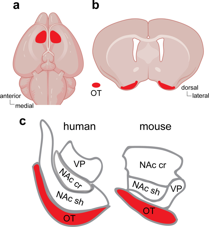
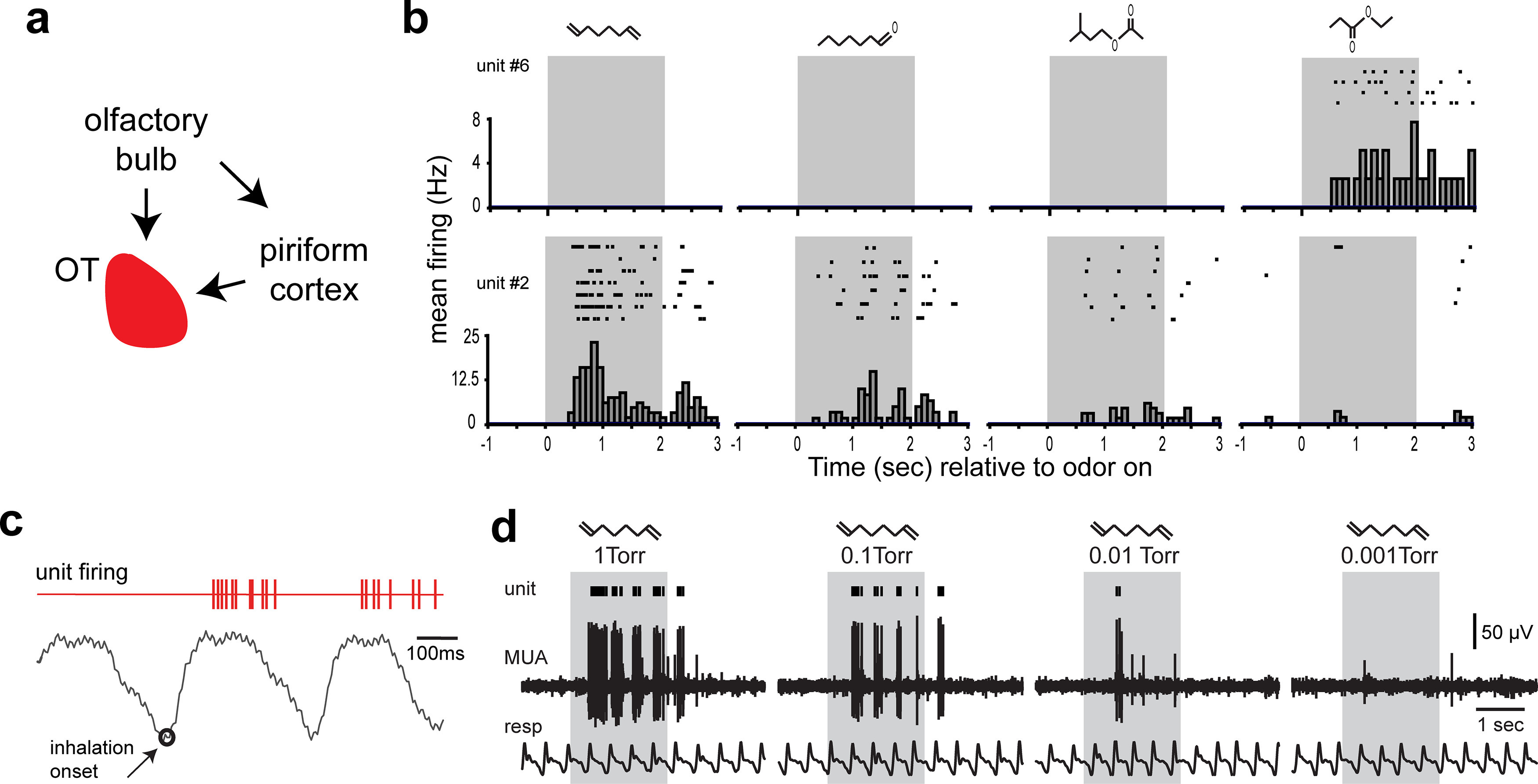
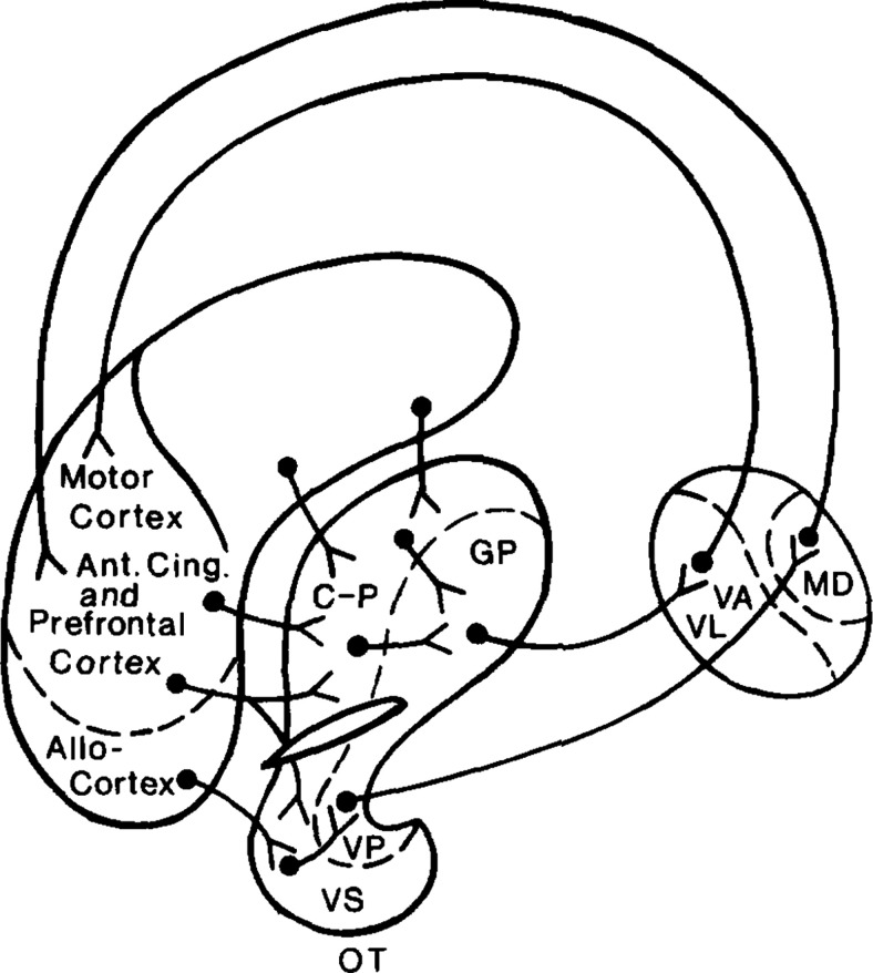
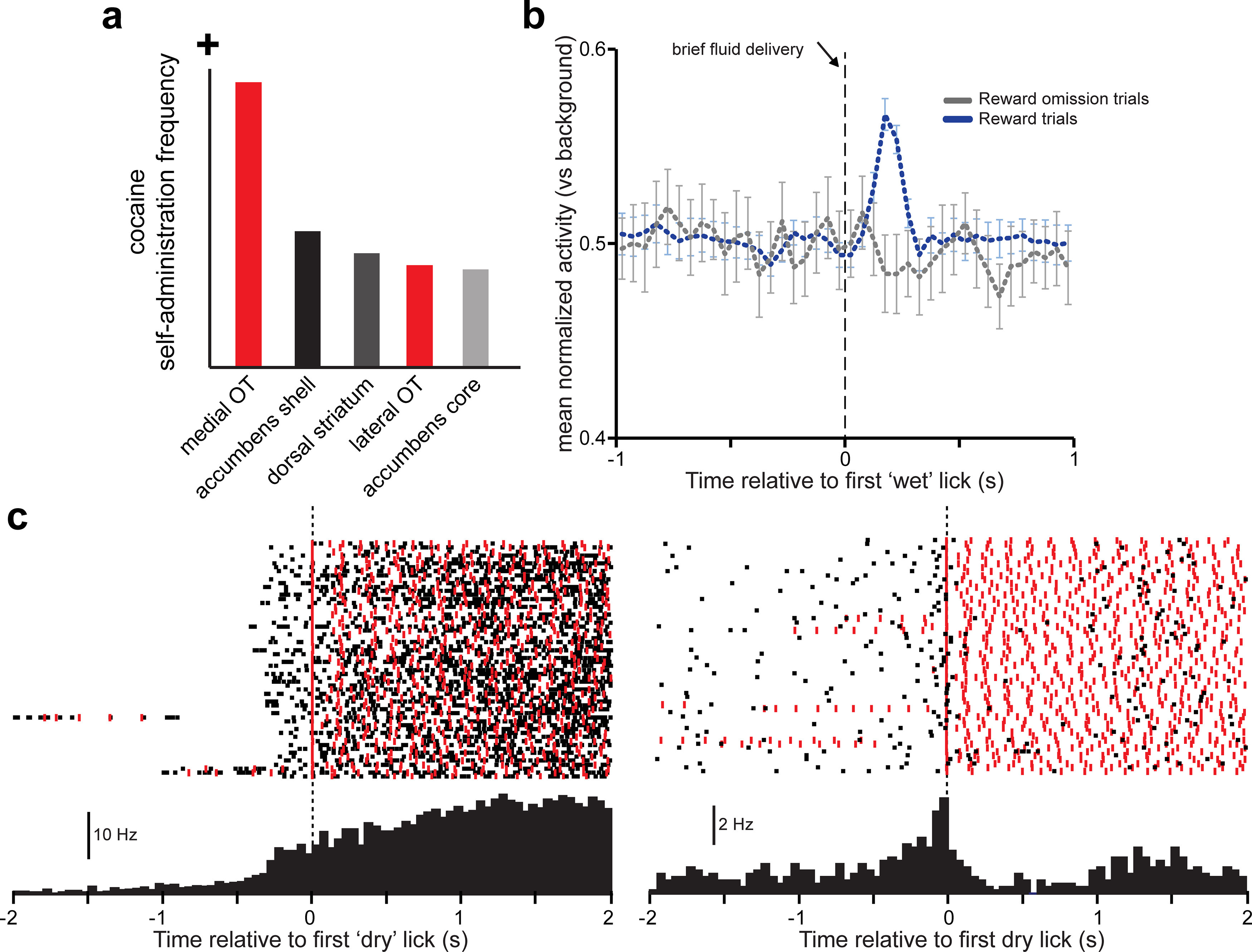
In the mid-19th century, a misconception was born, which understandably persists in the minds of many neuroscientists today. The eminent scientist Albert von Kölliker named a tubular-shaped piece of tissue found in the brains of all mammals studied to date, the tuberculum olfactorium - or what is commonly known as the olfactory tubercle (OT). In doing this, Kölliker ascribed "olfactory" functions and an "olfactory" purpose to the OT. The OT has since been classified as one of several olfactory cortices. However, further investigations of OT functions, especially over the last decade, have provided evidence for roles of the OT beyond olfaction, including in learning, motivated behaviors, and even seeking of psychoactive drugs. Indeed, research to date suggests caution in assigning the OT with a purely olfactory role. Here, I build on previous research to synthesize a model wherein the OT, which may be more appropriately termed the "tubular striatum" (TuS), is a neural system in which sensory information derived from an organism's experiences is integrated with information about its motivational states to guide affective and behavioral responses.


Figures

Figure 1.
Figure 1.
Localization of the OT in mice and humans. Approximate areas of the OT in the mouse brain from both the inferior (a) and coronal (b) views. c, Illustration of the relative location of the OT in humans and mice in respect to the neighboring nucleus accumbens shell (NAc sh) and core (NAc cr) and the ventral pallidum (VP).
Figure 2.
Figure 2.
Odor input into the OT and the representation of odors by OT neurons. a, Schematic of the major sources of odor information into the OT, including from the olfactory bulb and piriform cortex, which receives direct input from the olfactory bulb to extend disynaptic input to the OT. Data from Gurdjian (1925), L. E. White (1965), Scott et al. (1980), and Schwob and Price (1984b). b, Example of odor-evoked activity in two OT single units from two separate urethane-anesthetized mice. Data are single-unit raster plots and peristimulus time histograms across multiple presentations with four different odorants (from left to right: 1,7-octadiene, ethyl propionate, heptanal, and isoamyl acetate). The top unit displays narrow tuning, responding to only one of the odorants, whereas the bottom unit responds to several odorants. Data adapted from Wesson and Wilson (2010). c, Example single-unit raster of spiking events (red) during odor presentation in relation to respiratory cycles, indicating the short latency until spiking from this unit relative to the onset of inhalation (open circle). Data adapted from Payton et al. (2012). d, Example multiunit OT trace (MUA) and single-unit raster plot (unit) along with respiration (resp) from a single recording in a urethane-anesthetized mouse showing reduction in spiking with decreased intensities of a single odor (1,7-octadiene). Whereas the 1 torr intensity odor elicited robust spiking during most inhalation events, the single unit in this example did not spike to the lowest intensity. Data adapted from Xia et al. (2015).
Figure 3.
Figure 3.
Lennart Heimer's original drawing, illustrating his concept that the ventral striatopallidum is integral with the basal ganglia's striato-pallidopthalamic loop. Reproduced with permission from Heimer et al. (1982). VS, Ventral striatum; VP, ventral pallidum; MD, medio-dorsal thalamus; VA, ventralis anterior complex of the thalamus; VL, ventralis lateralis complex of the thalamus; GP, globus pallidus; C-P, caudate putamen. While not indicated in this illustration, Heimer included the nucleus accumbens as part of the VS.
Figure 4.
Figure 4.
Evidence that the OT supports the reinforcing properties of psychoactive drugs and encodes rewards. a, Summary of results from the work of Ikemoto (2003), indicating the frequency of intracranial cocaine infusions into ventral striatum subregions (red represents OT) when rats were allowed to self-administer 200 mm cocaine. Rats more frequently self-administered cocaine into the medial OT than any other brain region tested. b, Population-averaged firing of OT single units (normalized) during the engagement of mice in a water-motivated task requiring them to lick in succession to obtain a fluid reward. Receipt of reward on the first lick (“wet” lick) evoked a brief bust of firing in the units; this was not observed on trials in which the reward was withheld (omission). c, Example activity of two single units from separate mice (black rasters across trials and peristimulus time histograms averaged across trials) recorded in the same paradigm as in b, but relative to the time the animal initiated the successive licks yet received no reinforcer (“dry” lick). Red ticks represent timing of licks. Both example units increased firing before the onset of the instrumental response to obtain the reward; and in the example to the right, the firing was not related to timing of licks. b, c, Adapted with permission from Gadziola and Wesson (2016).

References

    1. Agustín-Pavón C, Martínez-García F, Lanuza E (2014) Focal lesions within the ventral striato-pallidum abolish attraction for male chemosignals in female mice. Behav Brain Res 259:292–296. 10.1016/j.bbr.2013.11.020 - DOI - PubMed
    1. Alheid GF, Heimer L (1988) New perspectives in basal forebrain organization of special relevance for neuropsychiatric disorders: the striatopallidal, amygdaloid, and corticopetal components of substantia innominata. Neuroscience 27:1–39. 10.1016/0306-4522(88)90217-5 - DOI - PubMed
    1. Anderson AK, Christoff K, Stappen I, Panitz D, Ghahremani DG, Glover G, Gabrieli JD, Sobel N (2003) Dissociated neural representations of intensity and valence in human olfaction. Nat Neurosci 6:196–202. 10.1038/nn1001 - DOI - PubMed
    1. Cansler HL, Wright KN, Stetzik LA, Wesson DW (2020) Neurochemical organization of the ventral striatum's olfactory tubercle. J Neurochem 152:425–448. 10.1111/jnc.14919 - DOI - PMC - PubMed
    1. Carelli RM. (2002) The nucleus accumbens and reward: neurophysiological investigations in behaving animals. Behav Cogn Neurosci Rev 1:281–296. 10.1177/1534582302238338 - DOI - PubMed

Publication types

LinkOut - more resources