

Effects of early ketamine exposure on cerebral gray matter volume and functional connectivity
- PMID: 32968108
- PMCID: PMC7512006
- DOI: 10.1038/s41598-020-72320-z
Effects of early ketamine exposure on cerebral gray matter volume and functional connectivity
Abstract
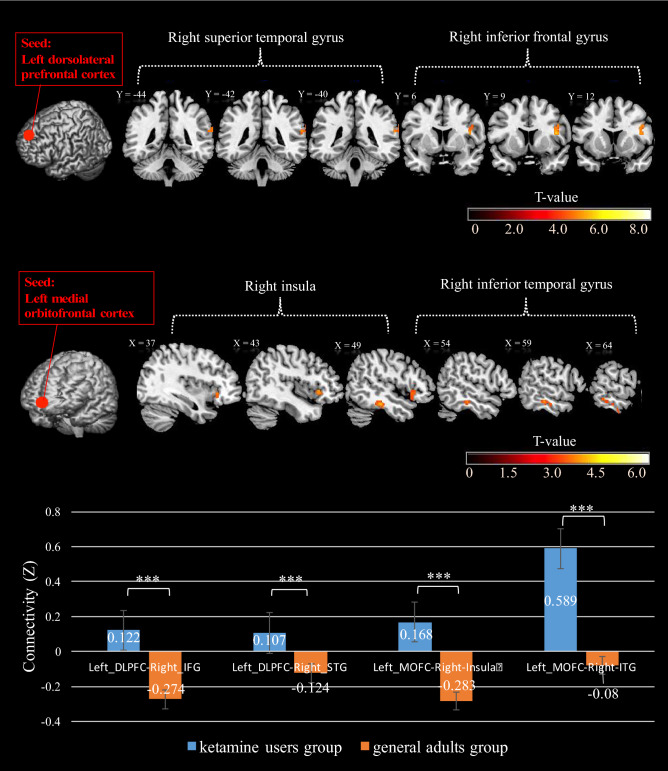
Ketamine has been used for medical purposes, most typically as an anesthetic, and recent studies support its use in the treatment of depression. However, ketamine tends to be abused by adolescents and young adults. In the current study, we examined the effects of early ketamine exposure on brain structure and function. We employed MRI to assess the effects of ketamine abuse on cerebral gray matter volume (GMV) and functional connectivity (FC) in 34 users and 19 non-users, employing covariates. Ketamine users were categorized as adolescent-onset and adult-onset based on when they were first exposed to ketamine. Imaging data were processed by published routines in SPM and AFNI. The results revealed lower GMV in the left precuneus in ketamine users, with a larger decrease in the adolescent-onset group. The results from a seed-based correlation analysis show that both ketamine groups had higher functional connectivity between left precuneus (seed) and right precuneus than the control group. Compared to controls, ketamine users showed decreased GMV in the right insula, left inferior parietal lobule, left dorsolateral prefrontal cortex/superior frontal gyrus, and left medial orbitofrontal cortex. These preliminary results characterize the effects of ketamine misuse on brain structure and function and highlight the influence of earlier exposure to ketamine on the development of the brain. The precuneus, a structure of central importance to cerebral functional organization, may be particularly vulnerable to the influences of early ketamine exposure. How these structural and functional brain changes may relate to the cognitive and affective deficits remains to be determined with a large cohort of participants.
Conflict of interest statement
The authors declare no competing interests.
Figures


Similar articles
-
Potential gray matter unpruned in adolescents and young adults dependent on dextromethorphan-containing cough syrups: evidence from cortical and subcortical study.Brain Imaging Behav. 2017 Oct;11(5):1470-1478. doi: 10.1007/s11682-016-9628-0. Brain Imaging Behav. 2017. PMID: 27738991
-
Common and distinct abnormal frontal-limbic system structural and functional patterns in patients with major depression and bipolar disorder.Neuroimage Clin. 2018 Jul 6;20:42-50. doi: 10.1016/j.nicl.2018.07.002. eCollection 2018. Neuroimage Clin. 2018. PMID: 30069426 Free PMC article.
-
Structural and functional brain abnormalities in schizophrenia: A cross-sectional study at different stages of the disease.Prog Neuropsychopharmacol Biol Psychiatry. 2018 Apr 20;83:27-32. doi: 10.1016/j.pnpbp.2017.12.017. Epub 2017 Dec 29. Prog Neuropsychopharmacol Biol Psychiatry. 2018. PMID: 29292241
-
Cortical and Subcortical Gray Matter Volume in Youths With Conduct Problems: A Meta-analysis.JAMA Psychiatry. 2016 Jan;73(1):64-72. doi: 10.1001/jamapsychiatry.2015.2423. JAMA Psychiatry. 2016. PMID: 26650724 Review.
-
Brain Changes Associated With Long-Term Ketamine Abuse, A Systematic Review.Front Neuroanat. 2022 Mar 18;16:795231. doi: 10.3389/fnana.2022.795231. eCollection 2022. Front Neuroanat. 2022. PMID: 35370568 Free PMC article.
Cited by
-
Adolescent substance use initiation and long-term neurobiological outcomes: insights, challenges and opportunities.Mol Psychiatry. 2024 Jul;29(7):2211-2222. doi: 10.1038/s41380-024-02471-2. Epub 2024 Feb 26. Mol Psychiatry. 2024. PMID: 38409597 Review.
-
Major challenges in youth psychopathology: treatment-resistant depression. A narrative review.Front Psychiatry. 2024 Jul 11;15:1417977. doi: 10.3389/fpsyt.2024.1417977. eCollection 2024. Front Psychiatry. 2024. PMID: 39056019 Free PMC article. Review.
-
Clinical and behavior characteristics of individuals who used ketamine.Sci Rep. 2022 Jan 17;12(1):801. doi: 10.1038/s41598-022-04832-9. Sci Rep. 2022. PMID: 35039593 Free PMC article.
-
Severe Encephalatrophy and Related Disorders From Long-Term Ketamine Abuse: A Case Report and Literature Review.Front Psychiatry. 2021 Oct 1;12:707326. doi: 10.3389/fpsyt.2021.707326. eCollection 2021. Front Psychiatry. 2021. PMID: 34658951 Free PMC article.
-
"K-Powder" Exposure during Adolescence Elicits Psychiatric Disturbances Associated with Oxidative Stress in Female Rats.Pharmaceuticals (Basel). 2022 Nov 9;15(11):1373. doi: 10.3390/ph15111373. Pharmaceuticals (Basel). 2022. PMID: 36355545 Free PMC article.
