

Structural analysis of a new carotenoid-binding protein: the C-terminal domain homolog of the OCP
- PMID: 32968135
- PMCID: PMC7512017
- DOI: 10.1038/s41598-020-72383-y
Structural analysis of a new carotenoid-binding protein: the C-terminal domain homolog of the OCP
Abstract
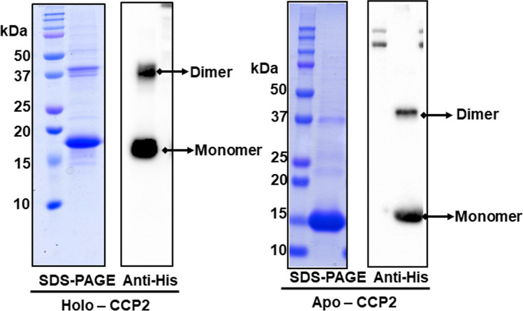
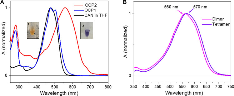
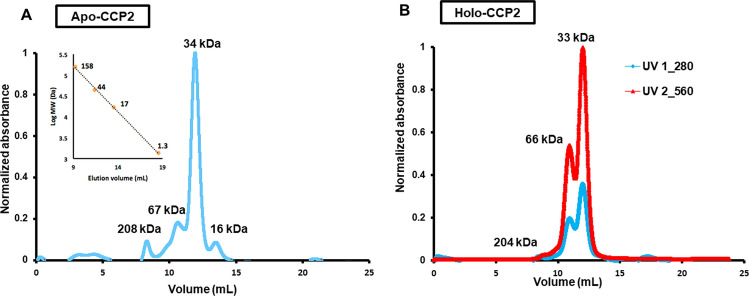
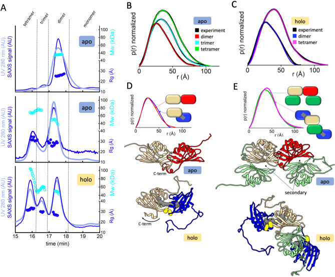
The Orange Carotenoid Protein (OCP) is a water-soluble protein that governs photoprotection in many cyanobacteria. The 35 kDa OCP is structurally and functionally modular, consisting of an N-terminal effector domain (NTD) and a C-terminal regulatory domain (CTD); a carotenoid spans the two domains. The CTD is a member of the ubiquitous Nuclear Transport Factor-2 (NTF2) superfamily (pfam02136). With the increasing availability of cyanobacterial genomes, bioinformatic analysis has revealed the existence of a new family of proteins, homologs to the CTD, the C-terminal domain-like carotenoid proteins (CCPs). Here we purify holo-CCP2 directly from cyanobacteria and establish that it natively binds canthaxanthin (CAN). We use small-angle X-ray scattering (SAXS) to characterize the structure of this carotenoprotein in two distinct oligomeric states. A single carotenoid molecule spans the two CCPs in the dimer. Our analysis with X-ray footprinting-mass spectrometry (XFMS) identifies critical residues for carotenoid binding that likely contribute to the extreme red shift (ca. 80 nm) of the absorption maximum of the carotenoid bound by the CCP2 dimer and a further 10 nm shift in the tetramer form. These data provide the first structural description of carotenoid binding by a protein consisting of only an NTF2 domain.
Conflict of interest statement
The authors declare no competing interests.
Figures
Similar articles
-
Synthetic OCP heterodimers are photoactive and recapitulate the fusion of two primitive carotenoproteins in the evolution of cyanobacterial photoprotection.Plant J. 2017 Aug;91(4):646-656. doi: 10.1111/tpj.13593. Epub 2017 Jun 12. Plant J. 2017. PMID: 28503830
-
Structural and spectroscopic characterization of HCP2.Biochim Biophys Acta Bioenerg. 2019 May 1;1860(5):414-424. doi: 10.1016/j.bbabio.2019.03.004. Epub 2019 Mar 15. Biochim Biophys Acta Bioenerg. 2019. PMID: 30880081
-
Assembly of photoactive orange carotenoid protein from its domains unravels a carotenoid shuttle mechanism.Photosynth Res. 2017 Sep;133(1-3):327-341. doi: 10.1007/s11120-017-0353-3. Epub 2017 Feb 17. Photosynth Res. 2017. PMID: 28213741
-
Structure and functions of Orange Carotenoid Protein homologs in cyanobacteria.Curr Opin Plant Biol. 2017 Jun;37:1-9. doi: 10.1016/j.pbi.2017.03.010. Epub 2017 Apr 6. Curr Opin Plant Biol. 2017. PMID: 28391046 Review.
-
Structure, function and evolution of the cyanobacterial orange carotenoid protein and its homologs.New Phytol. 2017 Aug;215(3):937-951. doi: 10.1111/nph.14670. Epub 2017 Jul 4. New Phytol. 2017. PMID: 28675536 Review.
Cited by
-
Heterologous expression of cyanobacterial Orange Carotenoid Protein (OCP2) as a soluble carrier of ketocarotenoids in Chlamydomonas reinhardtii.Algal Res. 2021 Mar 5;55:102255. doi: 10.1016/j.algal.2021.102255. eCollection 2021 May. Algal Res. 2021. PMID: 33777686 Free PMC article.
-
Recent Progress in Solution Structure Studies of Photosynthetic Proteins Using Small-Angle Scattering Methods.Molecules. 2023 Nov 3;28(21):7414. doi: 10.3390/molecules28217414. Molecules. 2023. PMID: 37959833 Free PMC article. Review.
-
Comparison of angiopoietin-like protein 3 and 4 reveals structural and mechanistic similarities.J Biol Chem. 2021 Jan-Jun;296:100312. doi: 10.1016/j.jbc.2021.100312. Epub 2021 Jan 20. J Biol Chem. 2021. PMID: 33482195 Free PMC article.
-
Enabling simultaneous photoluminescence spectroscopy and X-ray footprinting mass spectrometry to study protein conformation and interactions.Anal Methods. 2025 Feb 6;17(6):1214-1225. doi: 10.1039/d4ay01670j. Anal Methods. 2025. PMID: 39749913
-
Integrated Structural Studies for Elucidating Carotenoid-Protein Interactions.Adv Exp Med Biol. 2022;1371:1-10. doi: 10.1007/5584_2020_615. Adv Exp Med Biol. 2022. PMID: 33963527 Free PMC article.
References
-
- Esteban R, Moran JF, Becerril JMA, Garcia-Plazaola JI. Versatility of carotenoids: An integrated view on diversity, evolution, functional roles and environmental interactions. Environ. Exp. Bot. 2015;119:63–75. doi: 10.1016/j.envexpbot.2015.04.009. - DOI
