. 2020 Sep 23;10(1):15551.
doi: 10.1038/s41598-020-72755-4.

Evaluation of the neuroprotective effect of taurine in Alzheimer's disease using functional molecular imaging

Affiliations

Evaluation of the neuroprotective effect of taurine in Alzheimer's disease using functional molecular imaging

Se Jong Oh et al. Sci Rep. .

Abstract

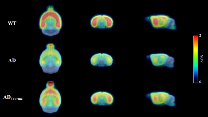
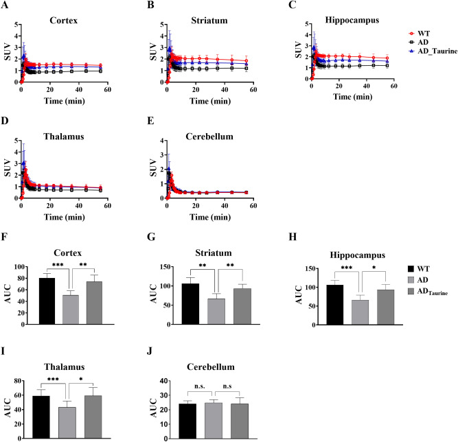
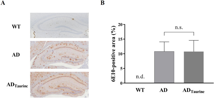
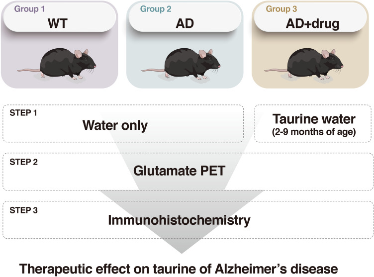
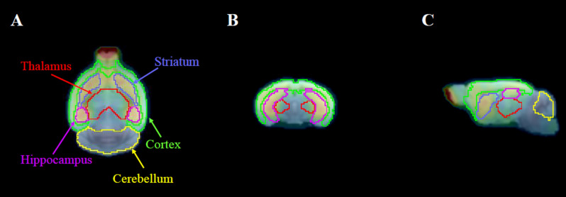
Alzheimer's disease (AD) is a chronic neurodegenerative disorder and the leading cause of dementia, but therapeutic treatment options are limited. Taurine has been reported to have neuroprotective properties against dementia, including AD. The present study aimed to investigate the treatment effect of taurine in AD mice by functional molecular imaging. To elucidate glutamate alterations by taurine, taurine was administered to 5xFAD transgenic mice from 2 months of age, known to apear amyloid deposition. Then, we performed glutamate positron emission tomography (PET) imaging studies for three groups (wild-type, AD, and taurine-treated AD, n = 5 in each group). As a result, brain uptake in the taurine-treated AD group was 31-40% higher than that in the AD group (cortex: 40%, p < 0.05; striatum: 32%, p < 0.01; hippocampus: 36%, p < 0.01; thalamus: 31%, p > 0.05) and 3-14% lower than that in the WT group (cortex: 10%, p > 0.05; striatum: 15%, p > 0.05; hippocampus: 14%, p > 0.05; thalamus: 3%, p > 0.05). However, we did not observe differences in Aβ pathology between the taurine-treated AD and AD groups in immunohistochemistry experiments. Our results reveal that although taurine treatment did not completely recover the glutamate system, it significantly increased metabolic glutamate receptor type 5 brain uptake. Therefore, taurine has therapeutic potential against AD.


Conflict of interest statement

The authors declare no competing interests.

Figures

Figure 1
Figure 1
Mean 18F-FPEB PET images (n = 5 for each group) between 30 and 60 min after injection in the three groups. All mean PET images were created using PMOD software (version 3.4). The AD group showed dramatically lower uptake than the WT group, and the ADTaurine group exhibited relatively higher uptake than the AD group. Images are shown scaled to the SUV.
Figure 2
Figure 2
Time-activity curves of the cortex, striatum, hippocampus, thalamus and cerebellum regions (AE). Brain uptake in the taurine-treated AD group was higher than that in the AD group but lower than that in the WT group. AUC values for the target regions (FJ). AUC values in the AD group were lower than the corresponding values in the WT group, but the ADTaurine group showed higher AUC values than the AD group. Data represent the mean values ± SD (n = 5). *p < 0.05, **p < 0.01, ***p < 0.001, n.s. = statistically nonsignificant difference. Mean values were calculated and statistical analysis was performed using Prism software (version 8).
Figure 3
Figure 3
Immunohistochemical staining of Aβ in the brains of AD and ADTaurine mice (A). The insets represent high-magnification images of the hippocampus. No morphological difference in Aβ was observed between the AD and ADTaurine groups. Quantification of Aβ deposition in the hippocampus (B). No quantitative differences were observed between the AD and ADTaurine groups. Values are presented as the mean ± SD, n.s. = statistically nonsignificant difference, n.d. = not detected. All statistical analysis was performed using Prism software (version 8).
Figure 4
Figure 4
Schematic of the study process.
Figure 5
Figure 5
Definition of VOIs in an AD mouse in the horizontal (A), coronal (B), and sagittal (C) planes. VOI applied PET images were produced using the PMOD fusion tool (version 3.4).

Similar articles

Cited by

References

    1. Tarawneh R, Holtzman DM. The clinical problem of symptomatic Alzheimer disease and mild cognitive impairment. Cold Spring Harb. Perspect. Med. 2012;2:1–16. doi: 10.1101/cshperspect.a006148. - DOI - PMC - PubMed
    1. Colvez A, Joël ME, Ponton-Sanchez A, Royer AC. Health status and work burden of Alzheimer patients’ informal caregivers: comparisons of five different care programs in the European Union. Health Policy. 2002;60:219–233. doi: 10.1016/S0168-8510(01)00215-9. - DOI - PubMed
    1. Halpern R, et al. Using electronic health records to estimate the prevalence of agitation in Alzheimer disease/dementia. Int. J. Geriatr. Psychiatry. 2019;34:420–431. doi: 10.1002/gps.5030. - DOI - PMC - PubMed
    1. Association A. 2018 Alzheimer’s disease facts and figures. Alzheimer’s Dement. 2018;14:367–429. doi: 10.1016/j.jalz.2018.02.001. - DOI
    1. Uddin MS, et al. Circadian and sleep dysfunction in Alzheimer’s disease. Ageing Res. Rev. 2020;60:1–2. doi: 10.1016/j.arr.2020.101046. - DOI - PubMed

Publication types

MeSH terms