. 2020 Oct;586(7827):145-150.
doi: 10.1038/s41586-020-2761-3. Epub 2020 Sep 23.

Synthetic group A streptogramin antibiotics that overcome Vat resistance

Affiliations

Synthetic group A streptogramin antibiotics that overcome Vat resistance

Qi Li et al. Nature. 2020 Oct.

Abstract

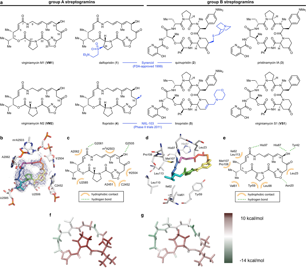
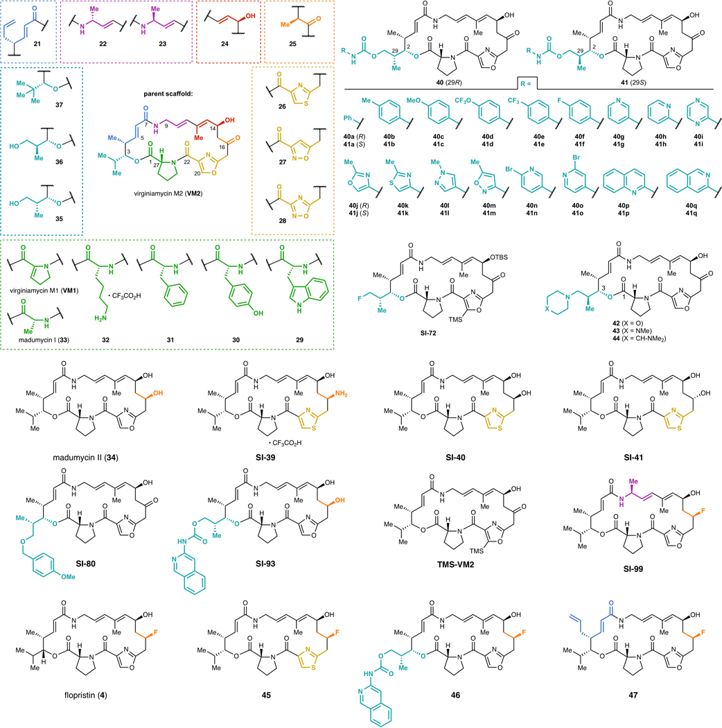
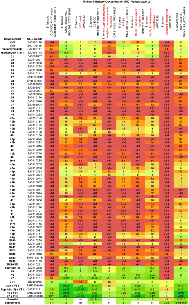
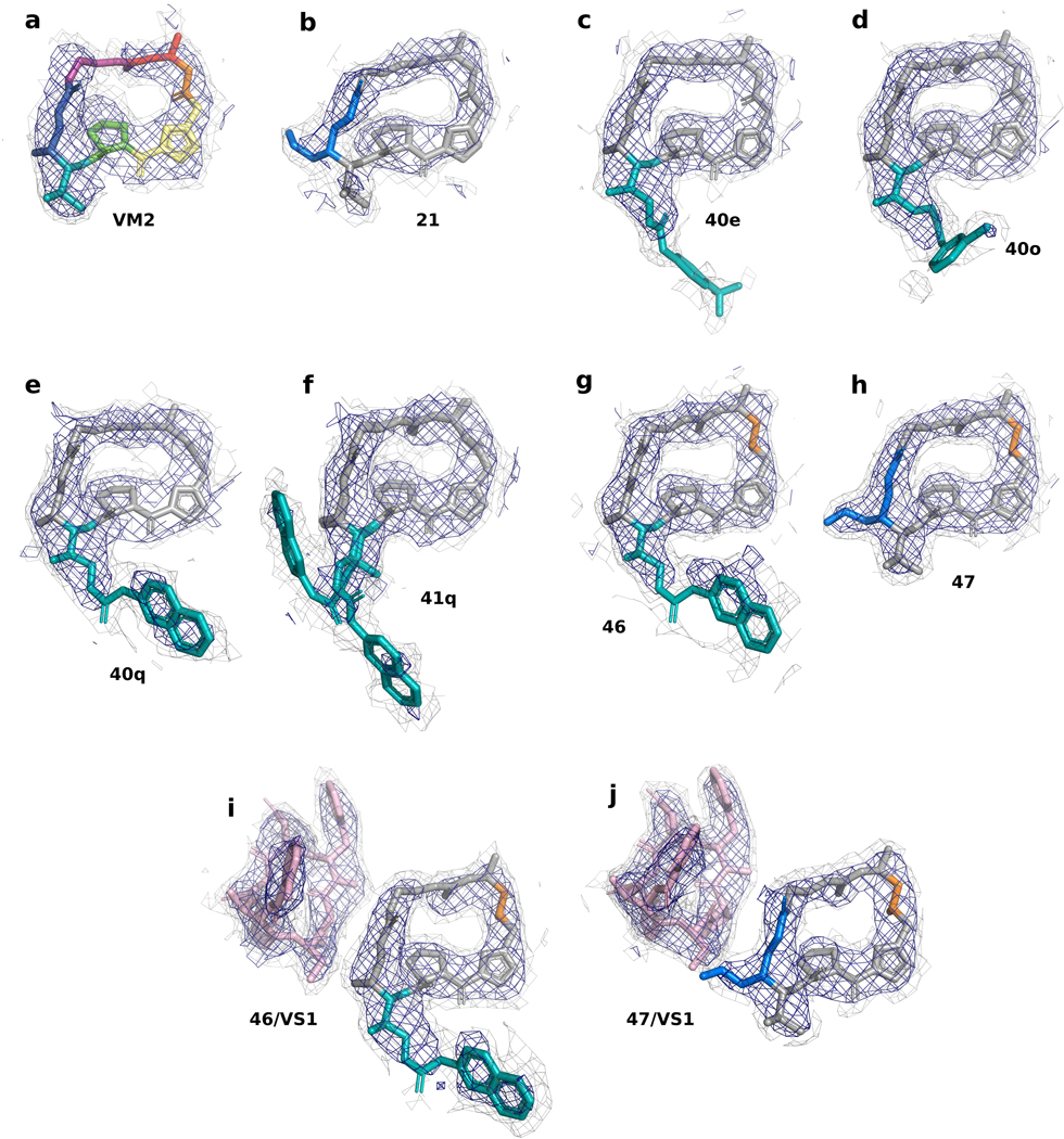
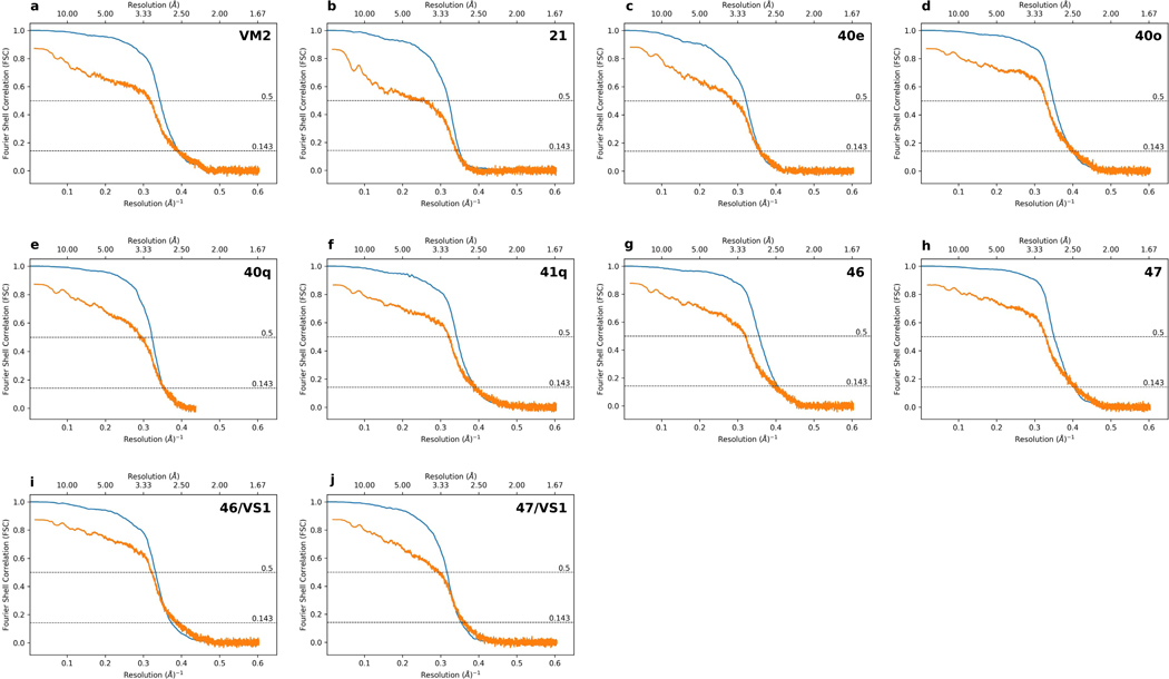
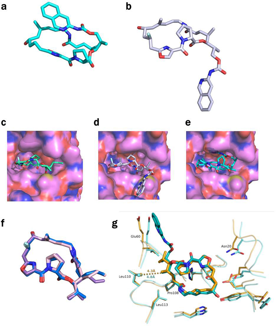
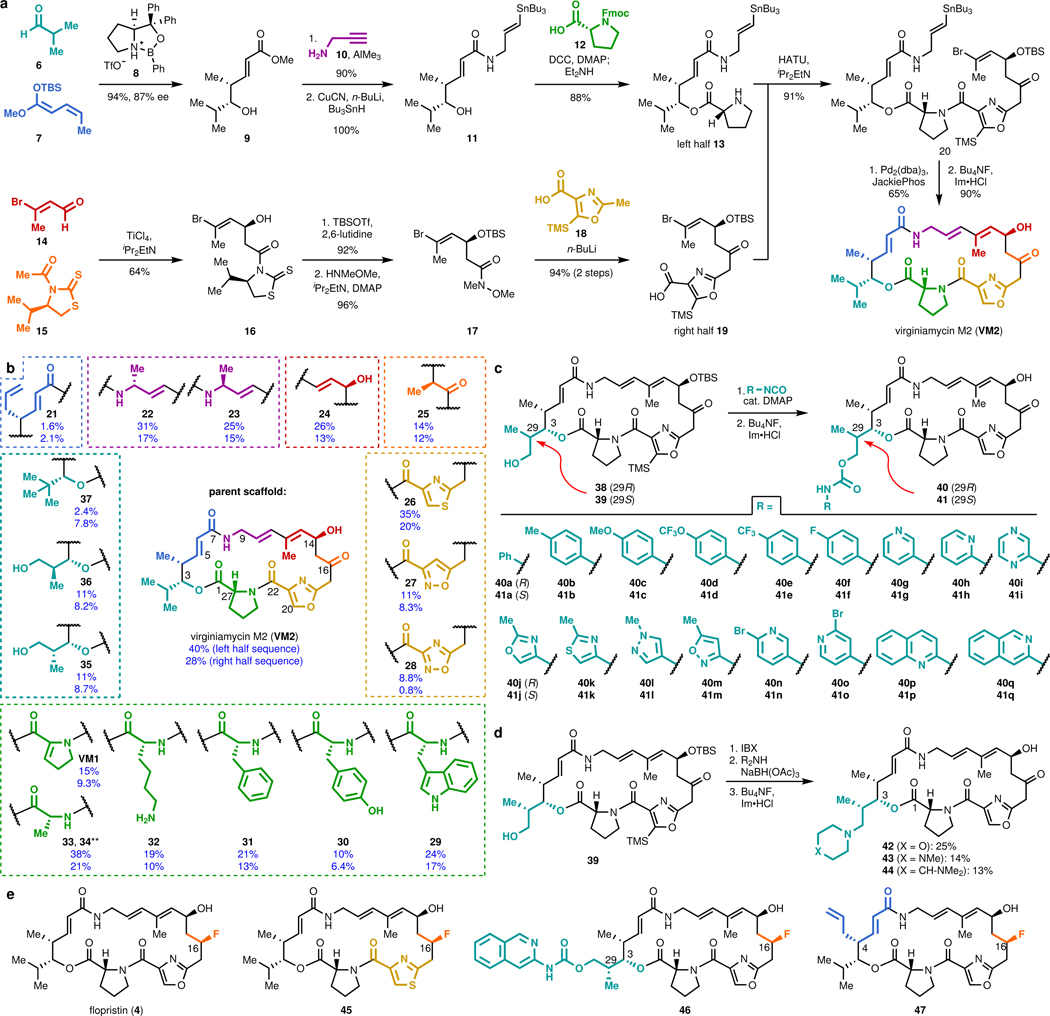
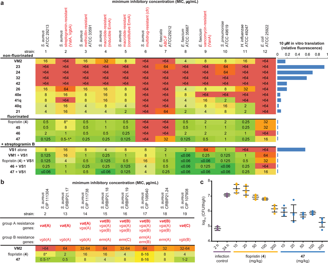
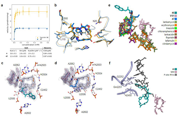
Natural products serve as chemical blueprints for most antibiotics in clinical use. The evolutionary process by which these molecules arise is inherently accompanied by the co-evolution of resistance mechanisms that shorten the clinical lifetime of any given class of antibiotics1. Virginiamycin acetyltransferase (Vat) enzymes are resistance proteins that provide protection against streptogramins2, potent antibiotics against Gram-positive bacteria that inhibit the bacterial ribosome3. Owing to the challenge of selectively modifying the chemically complex, 23-membered macrocyclic scaffold of group A streptogramins, analogues that overcome the resistance conferred by Vat enzymes have not been previously developed2. Here we report the design, synthesis, and antibacterial evaluation of group A streptogramin antibiotics with extensive structural variability. Using cryo-electron microscopy and forcefield-based refinement, we characterize the binding of eight analogues to the bacterial ribosome at high resolution, revealing binding interactions that extend into the peptidyl tRNA-binding site and towards synergistic binders that occupy the nascent peptide exit tunnel. One of these analogues has excellent activity against several streptogramin-resistant strains of Staphylococcus aureus, exhibits decreased rates of acetylation in vitro, and is effective at lowering bacterial load in a mouse model of infection. Our results demonstrate that the combination of rational design and modular chemical synthesis can revitalize classes of antibiotics that are limited by naturally arising resistance mechanisms.


Figures

Extended Data Figure 1 |
Extended Data Figure 1 |. Natural and semisynthetic streptogramins and their molecular mechanisms of action and resistance.
a, Selected natural and semisynthetic streptogramin analogs. Modifications installed by semisynthesis are highlighted in blue. b, 2.5-Å cryo-EM structure of VM2 bound to the 50S subunit of the E. coli ribosome. Coulomb potential density is contoured in dark blue at 4.0𝜎 and light gray at 1.0𝜎. Atom coloring of VM2 mirrors the building blocks used in its synthesis (see Figure 2). c, Binding interactions between VM2 and residues in the ribosomal binding site. d, X-ray crystal structure VM1 bound to the resistance protein VatA (PDB ID: 4HUS). e, Binding interactions between VM1 and VatA, highlighting the extensive hydrophobic interactions at C3-C6. Acetylation occurs at the C14 alcohol. f,g, Conformational energy of VM2 showing contributions on a per atom basis when refined with standard CIF-based restraints generated by phenix.eLBOW (f) and when refined with OPLS3e/VSGB2.1 force field (g). Color indicates low strain (green, −14 kcal/mol) up to high strain (red, 10 kcal/mol), with total conformational energy of 39.5 kcal/mol (f) and −88.3 (g). Hydrogens were added and optimized with fixed heavy atoms for the CIF-based refined conformation using prepwizard; the PHENIX-OPLS3e/VSGB2.1 refined conformation was taken as is. Energies were calculated using Prime and per atom contribution visualized using Maestro’s Prime Energy Visualization.
Extended Data Figure 2 -
Extended Data Figure 2 -. List of streptogramins tested for inhibitory activity
Fully synthetic group A streptogramins tested for inhibitory activity against 21 strains of bacteria (see Extended Data Figures 3 and 4).
Extended Data Figure 3 -
Inhibitory activity against Gram-positive organisms
Extended Data Figure 4 -
Inhibitory activity against Gram-negative organisms
Extended Data Figure 5 -
Extended Data Figure 5 -. CryoEM Density for all compounds bound to the E coli ribosome
a, 2.6-Å CryoEM structure of VM2 bound to the 50S subunit of the E. coli ribosome. Coulomb potential density is contoured in dark blue at 4.0𝜎 and light gray at 1.0𝜎 for entire figure. b, 2.8-Å CryoEM structure of 21 bound to the 50S subunit of the E. coli ribosome. c, 2.8-Å CryoEM structure of 40e bound to the 50S subunit of the E. coli ribosome. d, 2.5-Å CryoEM structure of 40o bound to the 50S subunit of the E. coli ribosome. e, 2.8-Å CryoEM structure of 40q bound to the 50S subunit of the E. coli ribosome. f, 2.6-Å CryoEM structure of 41q bound to the 50S subunit of the E. coli ribosome. g, 2.5-Å CryoEM structure of 46 bound to the 50S subunit of the E. coli ribosome. h, 2.5-Å CryoEM structure of 47 bound to the 50S subunit of the E. coli ribosome. i, 2.7-Å CryoEM structure of 46/VS1 bound to the 50S subunit of the E. coli ribosome. j, 2.8-Å CryoEM structure of 47/VS1 bound to the 50S subunit of the E. coli ribosome.
Extended Data 6 -
Extended Data 6 -. Gold Standard and Map to Model Fourier Shell Correlation plots
a-j, The particle Fourier Shell Correlation (FSC) curves for reconstructions obtained by cisTEM using a molecular weight of 1.8 MDa are shown in blue with unmasked Map-Model FSC curves obtained from phenix.mtriage shown in orange. Dashed lines indicate FSC of 0.143 for estimating Gold Standard resolution and FSC of 0.5 for estimating Map-Model resolution.
Extended Data Figure 7 -
Extended Data Figure 7 -. Conformations of 46 and 47 in the ribosome and in VatA
a, The conformation of 46 minimized by QM methods in low dielectric, shows how the isoquinoline side chain packs over the macrocycle. b, In contrast, the ribosome-bound conformations of 46 determined by CryoEM show that the side chain extends away from the macrocycle due to interactions formed in the binding site. c, Model of 47 in the conformation bound to the ribosome modeled into the active site of VatA (shown in surface). d, Model of 46 in the conformation bound to the ribosome modeled into the active site of VatA. e, Low energy model of 46 modeled into the active site of VatA. f, Overlay of VatA-bound (marine), ribosome-bound (violet), and ribosome with VS1-bound (light pink) conformations of 47. g, X-ray crystal structures of VM1 bound to VatA (PDB ID: 4HUS, 2.4 Å) and 46 bound to VatA at 2.8-Å resolution.
Figure 1 |
Figure 1 |. Modular synthesis enables access to >60 fully synthetic group A streptogramins.
a, Convergent route to VM2 from seven building blocks. b, Eighteen group A streptogramins accessed by building block variation. The fragments displayed in the dashed boxes represent the structural variability compared to the parent scaffold (VM2). Overall yields for the synthesis of each analog for the left half sequence (top number) and for the right half sequence (bottom number) are displayed. **Instead of a ketone, madumycin II (34) contains the following substitution at C16: ɑ-H, β-OH. c, Access to 34 analogs (17 in each diastereomeric series) with C3 side chain variability by means of carbamate formation followed by desilylation. d, Synthesis of tertiary-amine-containing analogs by oxidation and reductive amination. e, C16-fluorinated analogs.
Figure 2 |
Figure 2 |. Antibiotic activity and in vivo efficacy of selected group A streptogramins.
a, MIC values for selected analogs against an expanded panel of pathogens. The bars to the right display in vitro translation that occurs in the presence of 10 μM of each analog (relative to DMSO). b, MIC values against eight clinical isolates of S. aureus with Vat resistance genes. The (*) indicates MIC values that were obtained in technical triplicate and biological triplicate. c, A murine thigh model of infection with S. aureus CIP 111304 (n=5 biologically independent animals per group, with the exception of the 2 h infection control where n=4 per group, examined over one experiment). Each animal is individually plotted, the center line is the mean, and the upper and lower whiskers bound the standard deviation from the mean. For detailed statistical analysis, see Extended Data Table 3.
Figure 3 |
Figure 3 |. In vitro acetylation, VatA binding, and ribosome binding of highly active analogs.
a, Summary of VatA acetylation kinetics and in vitro inhibition of the E. coli ribosome by 4 and 47. Error bars represent standard deviations of the mean (3 technical replicates). For detailed statistical analysis, see Extended Data Table 3. b, X-ray crystal structures of VM1 bound to VatA (PDB ID: 4HUS, 2.4 Å) and 47 bound to VatA at 3.2 Å resolution. Distances shown are measured between carbons of the C4 extension of 47 and Leu110 in the VM1-bound VatA structure (in orange dashes, 2.1 Å) and in the 47-bound VatA structure (in marine dashes, 3.6 Å). c, 2.7-Å cryo-EM Coulomb potential density map (contoured in dark blue at 4.0𝜎 and light gray at 1.0𝜎) for ribosomes bound to 46 and VS1. d, 2.8-Å cryo-EM Coulomb potential density map for ribosomes bound to 47 and VS1. e, An overlay of known PTC-site antibiotics shows how the side chain of 46 and the extension of 47 occupy areas distinct to previously characterized antibiotics. f, Overlay of P-site tRNA (dark gray, PDB ID: 1VY4) with the cryo-EM structure of ribosome-bound 46 reveals that the sidechain extends into the P-site and mimics the terminal adenosine (A2450) of the tRNA.

Comment in

References

    1. Wright PM, Seiple IB & Myers AG The evolving role of chemical synthesis in antibacterial drug discovery. Angew. Chem. Int. Ed Engl 53, 8840–8869 (2014). - PMC - PubMed
    1. Stogios PJ et al. Potential for reduction of streptogramin A resistance revealed by structural analysis of acetyltransferase VatA. Antimicrob. Agents Chemother 58, 7083–7092 (2014). - PMC - PubMed
    1. Vazquez D. The Streptogramin Family of Antibiotics in Mechanism of Action (eds. Gottlieb D. & Shaw PD) 387–403 (Springer Berlin; Heidelberg, 1967).
    1. Waglechner N. & Wright GD Antibiotic resistance: it’s bad, but why isn’t it worse? BMC Biol. 15, 84 (2017). - PMC - PubMed
    1. Seiple IB et al. A platform for the discovery of new macrolide antibiotics. Nature 533, 338–345 (2016). - PMC - PubMed

Extended Data References

    1. Tropea JE, Cherry S. & Waugh DS Expression and purification of soluble His(6)-tagged TEV protease. Methods Mol. Biol 498, 297–307 (2009). - PubMed
    1. Kuhn ML, Majorek KA, Minor W. & Anderson WF Broad-substrate screen as a tool to identify substrates for bacterial Gcn5-related N-acetyltransferases with unknown substrate specificity. Protein Sci. 22, 222–230 (2013). - PMC - PubMed
    1. Winter G. xia2: an expert system for macromolecular crystallography data reduction. Journal of Applied Crystallography vol. 43 186–190 (2010).
    1. Kabsch W. XDS. Acta Crystallogr. D Biol. Crystallogr 66, 125–132 (2010). - PMC - PubMed
    1. Liebschner D. et al. Macromolecular structure determination using X-rays, neutrons and electrons: recent developments in Phenix. Acta Crystallogr D Struct Biol 75, 861–877 (2019). - PMC - PubMed

Publication types

MeSH terms