

A nomogram based on glycomic biomarkers in serum and clinicopathological characteristics for evaluating the risk of peritoneal metastasis in gastric cancer
- PMID: 32968368
- PMCID: PMC7501696
- DOI: 10.1186/s12014-020-09297-4
A nomogram based on glycomic biomarkers in serum and clinicopathological characteristics for evaluating the risk of peritoneal metastasis in gastric cancer
Abstract
Background: Peritoneal metastasis (PM) in gastric cancer (GC) remains an untreatable disease, and is difficult to diagnose preoperatively. Here, we aim to establish a novel prediction model.
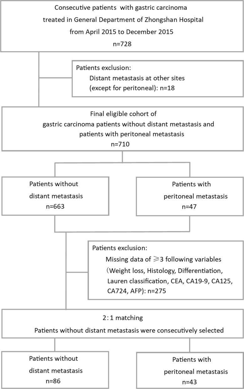
Methods: The clinicopathologic characteristics of a cohort that included 86 non-metastatic GC patients and 43 PMGC patients from Zhongshan Hospital were retrospectively analysed to identify PM associated variables. Additionally, mass spectrometry and glycomic analysis were applied in the same cohort to find glycomic biomarkers in serum for the diagnosis of PM. A nomogram was established based on the associations between potential risk variables and PM.
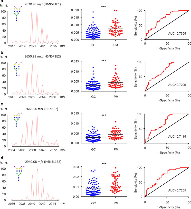
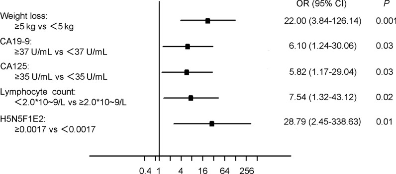
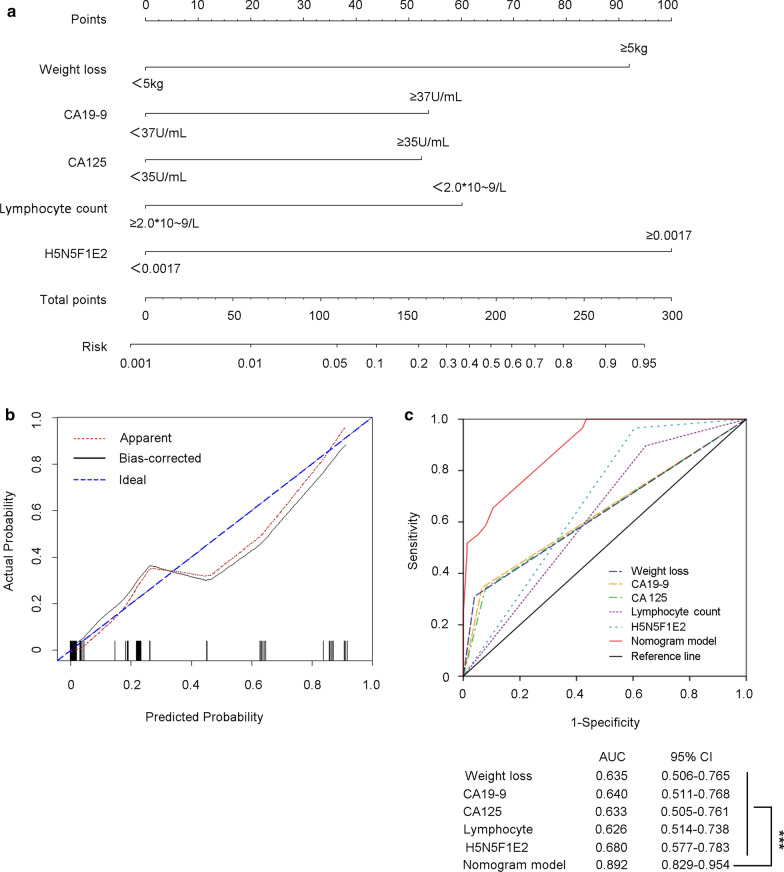
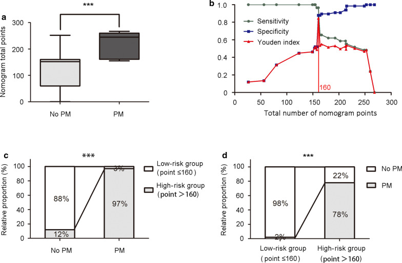
Results: Overexpression of 4 N-glycans (H6N5L1E1: m/z 2620.93; H5N5F1E2: m/z 2650.98; H6N5E2, m/z 2666.96; H6N5L1E2, m/z 2940.08); weight loss ≥ 5 kg; tumour size ≥ 3 cm; signet ring cell or mucinous adenocarcinoma histology type; poor differentiation; diffuse or mixed Lauren classification; increased CA19-9, CA125, and CA724 levels; decreased lymphocyte count, haemoglobin, albumin, and pre-albumin levels were identified to be associated with PM. A nomogram that integrated with five independent risk factors (weight loss ≥ 5 kg, CA19-9 ≥ 37 U/mL, CA125 ≥ 35 U/mL, lymphocyte count < 2.0 * 10 ~ 9/L, and H5N5F1E2 expression ≥ 0.0017) achieved a good performance for diagnosis (AUC: 0.892, 95% CI 0.829-0.954). When 160 was set as the cut-off threshold value, the proposed nomogram represented a perfectly discriminating power for both sensitivity (0.97) and specificity (0.88).
Conclusions: The nomogram achieved an individualized assessment of the risk of PM in GC patients; thus, the nomogram could be used to assist clinical decision-making before surgery.
Keywords: Gastric cancer; Glycomic analysis; MALDI-TOF–MS; Nomogram; Peritoneal metastasis.
© The Author(s) 2020.
Conflict of interest statement
Competing interestThe authors declare that they have no conflict of interest.
Figures
Similar articles
-
[Establishment of risk evaluation model of peritoneal metastasis in gastric cancer and its predictive value].Zhonghua Wei Chang Wai Ke Za Zhi. 2017 Jan 25;20(1):47-52. Zhonghua Wei Chang Wai Ke Za Zhi. 2017. PMID: 28105619 Chinese.
-
Practical nomogram based on comprehensive CT texture analysis to preoperatively predict peritoneal occult metastasis of gastric cancer patients.Front Oncol. 2022 Dec 1;12:882584. doi: 10.3389/fonc.2022.882584. eCollection 2022. Front Oncol. 2022. PMID: 36531010 Free PMC article.
-
Application of Circulating Tumor Cells and Interleukin-6 in Preoperative Prediction of Peritoneal Metastasis of Advanced Gastric Cancer.J Inflamm Res. 2023 Jul 20;16:3033-3047. doi: 10.2147/JIR.S414786. eCollection 2023. J Inflamm Res. 2023. PMID: 37497064 Free PMC article.
-
[Establishment and validation of a predictive nomogram model for advanced gastric cancer with perineural invasion].Zhonghua Wei Chang Wai Ke Za Zhi. 2020 Nov 25;23(11):1059-1066. doi: 10.3760/cma.j.cn.441530-20200103-00004. Zhonghua Wei Chang Wai Ke Za Zhi. 2020. PMID: 33212554 Chinese.
-
Clinical Significance of Serum CA125, CA19-9, CA72-4, and Fibrinogen-to-Lymphocyte Ratio in Gastric Cancer With Peritoneal Dissemination.Front Oncol. 2019 Nov 5;9:1159. doi: 10.3389/fonc.2019.01159. eCollection 2019. Front Oncol. 2019. PMID: 31750248 Free PMC article.
Cited by
-
LMOD1, an oncogene associated with Lauren classification, regulates the metastasis of gastric cancer cells through the FAK-AKT/mTOR pathway.BMC Cancer. 2022 Apr 30;22(1):474. doi: 10.1186/s12885-022-09541-0. BMC Cancer. 2022. PMID: 35488236 Free PMC article.
-
Application and progress of nomograms in gastric cancer.Front Med (Lausanne). 2025 Jan 29;12:1510742. doi: 10.3389/fmed.2025.1510742. eCollection 2025. Front Med (Lausanne). 2025. PMID: 39944483 Free PMC article. Review.
-
Intraoperative Pathological Evaluation of Suspicious Peritoneal Nodules for Surgical Decision-making in Gastric Cancer.J Gastrointest Surg. 2023 Aug;27(8):1545-1559. doi: 10.1007/s11605-023-05671-3. Epub 2023 Apr 14. J Gastrointest Surg. 2023. PMID: 37059962
-
Predicting peritoneal carcinomatosis of gastric cancer: A simple model to exempt low-risk patients from unnecessary staging laparoscopy.Front Surg. 2022 Jul 21;9:916001. doi: 10.3389/fsurg.2022.916001. eCollection 2022. Front Surg. 2022. PMID: 35937608 Free PMC article.
-
Novel nomogram and risk stratification for peritoneal recurrence after curative resection in gastric cancer.Sci Rep. 2024 Aug 17;14(1):19103. doi: 10.1038/s41598-024-70349-y. Sci Rep. 2024. PMID: 39154083 Free PMC article.
References
-
- Siegel RL, Miller KD, Jemal A. Cancer statistics, 2017. CA Cancer J Clin. 2017;67:7–30. - PubMed
-
- Emoto S, Ishigami H, Yamashita H, Yamaguchi H, Kaisaki S, Kitayama J. Clinical significance of CA125 and CA72-4 in gastric cancer with peritoneal dissemination. Gastric Cancer. 2012;15:154–161. - PubMed
-
- Kikuchi H, Kamiya K, Hiramatsu Y, Miyazaki S, Yamamoto M, Ohta M, Baba S, Konno H. Laparoscopic narrow-band imaging for the diagnosis of peritoneal metastasis in gastric cancer. Ann Surg Oncol. 2014;21:3954–3962. - PubMed
-
- Thomassen I, van Gestel YR, van Ramshorst B, Luyer MD, Bosscha K, Nienhuijs SW, Lemmens VE, de Hingh IH. Peritoneal carcinomatosis of gastric origin: a population-based study on incidence, survival and risk factors. Int J Cancer. 2014;134:622–628. - PubMed
LinkOut - more resources
Full Text Sources
Research Materials
Miscellaneous