

Ephrin B2 mediates high glucose induced endothelial-to-mesenchymal transition in human aortic endothelial cells
- PMID: 32968633
- PMCID: PMC7487366
- DOI: 10.21037/cdt-20-299
Ephrin B2 mediates high glucose induced endothelial-to-mesenchymal transition in human aortic endothelial cells
Abstract
Background: Previous study revealed that high glucose (HG) induced endothelial cell (EC) damage via endothelial-to-mesenchymal transition (EndMT). Recent studies suggested the role of Ephrin B2 in mediate ECs damage. However, the underlying mechanism remains unclear. The aim of the present study was to investigate whether Ephrin B2 mediates HG-induced EndMT in human aortic ECs (HAECs) and to determine the possible downstream signaling effector.
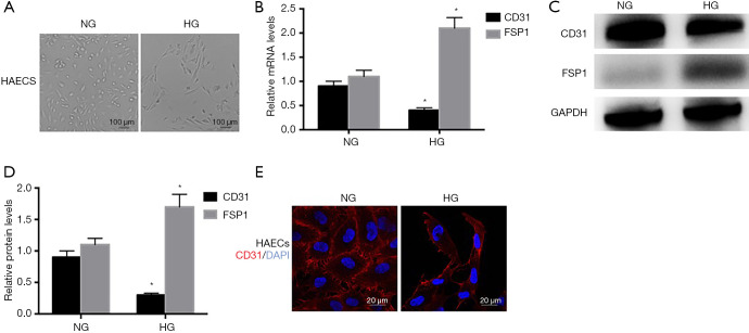
Methods: Primary HAECs were exposed to normal glucose (NG, 5.5 mM), HG (30 mM) and HG+Ephrin B2 small interfering RNA (siRNA), respectively. The pathological changes were investigated by light microscope and confocal microscopy. To study the effects of focal adhesion kinase (FAK) activation on Ephrin B2 in HAECs, cells were incubated with FAK siRNA in HG group. The expression of EndMT-related markers (CD31 and FSP1), Ephrin B2 and FAK were detected by qRT-PCR and western blot.
Results: The results showed that HG significantly inhibited the expression of CD31 and increased FSP1 compared with NG group. Moreover, Ephrin B2 was increased after HG incubation. Ephrin B2 siRNA attenuated HG-induced expression of EndMT-related markers. Furthermore, HG increased the expression of FAK and phosphorylated FAK (pho-FAK) in HAECs. In contrast, blocking Ephrin B2 could partially attenuate HG-induced FAK activation. And FAK siRNA further inhibited the EndMT-related markers in HAECs treated with HG.
Conclusions: HG-induced EndMT in HAECs might be partially mediated by Ephrin B2 and the downstream FAK pathway.
Keywords: Ephrin B2; endothelial-to-mesenchymal transition (EndMT); focal adhesion kinase (FAK); high glucose (HG); human aortic endothelial cell (HAEC).
2020 Cardiovascular Diagnosis and Therapy. All rights reserved.
Conflict of interest statement
Conflicts of Interest: All authors have completed the ICMJE uniform disclosure form (available at http://dx.doi.org/10.21037/cdt-20-299). The authors have no conflicts of interest to declare.
Figures


Similar articles
-
Calcium-Sensing Receptor Participates in High Glucose-Induced EndMT in Primary Human Aortic Endothelial Cells.Front Physiol. 2021 Jan 15;11:629542. doi: 10.3389/fphys.2020.629542. eCollection 2020. Front Physiol. 2021. PMID: 33519531 Free PMC article.
-
Interleukin-1β mediates high glucose induced phenotypic transition in human aortic endothelial cells.Cardiovasc Diabetol. 2016 Mar 5;15:42. doi: 10.1186/s12933-016-0358-9. Cardiovasc Diabetol. 2016. PMID: 26944557 Free PMC article.
-
High glucose mediates endothelial-to-chondrocyte transition in human aortic endothelial cells.Cardiovasc Diabetol. 2012 Sep 22;11:113. doi: 10.1186/1475-2840-11-113. Cardiovasc Diabetol. 2012. PMID: 22998723 Free PMC article.
-
Angiotensin II mediates the high-glucose-induced endothelial-to-mesenchymal transition in human aortic endothelial cells.Cardiovasc Diabetol. 2010 Jul 27;9:31. doi: 10.1186/1475-2840-9-31. Cardiovasc Diabetol. 2010. PMID: 20663195 Free PMC article.
-
Role of endothelial cells in vascular calcification.Front Cardiovasc Med. 2022 Jul 19;9:895005. doi: 10.3389/fcvm.2022.895005. eCollection 2022. Front Cardiovasc Med. 2022. PMID: 35928939 Free PMC article. Review.
Cited by
-
Aortic valve disease in diabetes: Molecular mechanisms and novel therapies.J Cell Mol Med. 2021 Oct;25(20):9483-9495. doi: 10.1111/jcmm.16937. Epub 2021 Sep 24. J Cell Mol Med. 2021. PMID: 34561944 Free PMC article. Review.
-
Nintedanib Inhibits Endothelial Mesenchymal Transition in Bleomycin-Induced Pulmonary Fibrosis via Focal Adhesion Kinase Activity Reduction.Int J Mol Sci. 2022 Jul 25;23(15):8193. doi: 10.3390/ijms23158193. Int J Mol Sci. 2022. PMID: 35897764 Free PMC article.
-
Calcium-Sensing Receptor Participates in High Glucose-Induced EndMT in Primary Human Aortic Endothelial Cells.Front Physiol. 2021 Jan 15;11:629542. doi: 10.3389/fphys.2020.629542. eCollection 2020. Front Physiol. 2021. PMID: 33519531 Free PMC article.
-
High Glucose Induced Changes in Human VEC Phenotype in a 3D Hydrogel Derived From Cell-Free Native Aortic Root.Front Cardiovasc Med. 2021 Aug 12;8:714573. doi: 10.3389/fcvm.2021.714573. eCollection 2021. Front Cardiovasc Med. 2021. PMID: 34458339 Free PMC article.
-
EphrinB2 alleviates tubulointerstitial fibrosis in diabetic kidney disease.J Transl Med. 2025 Jul 24;23(1):821. doi: 10.1186/s12967-025-06852-1. J Transl Med. 2025. PMID: 40707969 Free PMC article.
References
LinkOut - more resources
Full Text Sources
Research Materials
Miscellaneous