

Contrast imaging ultrasound for the detection and characterization of carotid vulnerable plaque
- PMID: 32968654
- PMCID: PMC7487408
- DOI: 10.21037/cdt.2020.01.08
Contrast imaging ultrasound for the detection and characterization of carotid vulnerable plaque
Abstract
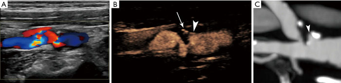
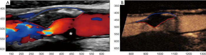
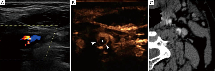
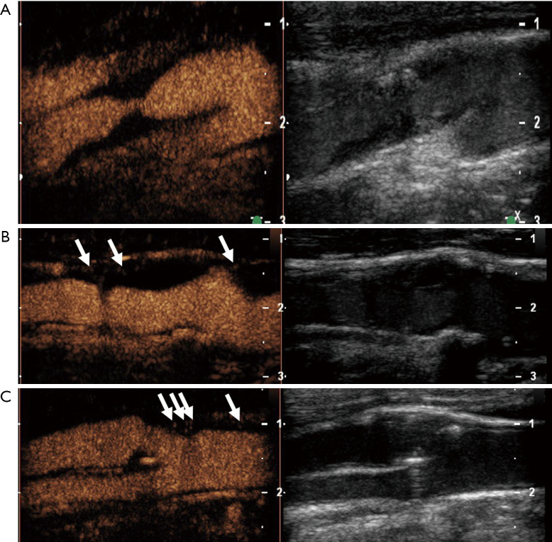
Not only the degree of luminal narrowing but also the plaque morphology and composition play an important role in risk stratification of carotid atherosclerotic lesions. During the last few years, carotid contrast-enhanced ultrasound (CEUS) has emerged as a valuable imaging tool to assess such vulnerable carotid plaques. This review article discussed the use of CEUS for the detection of carotid plaque irregularities and ulcerations as well as the quantification of intraplaque neovascularization and its correlation with histology and inflammatory biomarkers. Apart from evaluating for markers of vulnerable carotid plaques, CEUS enhancement is directly associated with past cerebrovascular events. More importantly, preliminary evidence has shown that CEUS could be used to predict future cerebrovascular and cardiovascular events. Despite the progress in CEUS imaging for carotid atherosclerotic disease, past studies still suffer from the retrospective nature, small sample size, and a lack of matched, well controlled prospective studies. In the future, large multi-center prospective studies addressing the relationship between CEUS findings and patient clinical outcomes in carotid atherosclerotic disease are warranted.
Keywords: Carotid plaque; contrast-enhanced ultrasound (CEUS); neovascularization; ulceration; ultrasound.
2020 Cardiovascular Diagnosis and Therapy. All rights reserved.
Conflict of interest statement
Conflicts of Interest: All authors have completed the ICMJE uniform disclosure form (available at http://dx.doi.org/10.21037/cdt.2020.01.08). The series “Advanced Imaging in The Diagnosis of Cardiovascular Diseases” was commissioned by the editorial office without any funding or sponsorship. SP and DS serve as the unpaid editorial board members of Cardiovascular Diagnosis and Therapy from July 2019 to June 2021. DS reports grants from Bracco SA, outside the submitted work. PSS reports personal fees from Bracco SA, outside the submitted work. The authors have no other conflicts of interest to declare.
Figures
Similar articles
-
Contrast-Enhanced Ultrasound Feasibility in Assessing Carotid Plaque Vulnerability-Narrative Review.J Clin Med. 2023 Oct 9;12(19):6416. doi: 10.3390/jcm12196416. J Clin Med. 2023. PMID: 37835061 Free PMC article. Review.
-
Contrast-Enhanced Ultrasound to Assess Carotid Intraplaque Neovascularization.Ultrasound Med Biol. 2020 Mar;46(3):466-478. doi: 10.1016/j.ultrasmedbio.2019.10.020. Epub 2019 Nov 29. Ultrasound Med Biol. 2020. PMID: 31791553
-
Contrast Enhanced Ultrasound (CEUS) Is Not Able to Identify Vulnerable Plaques in Asymptomatic Carotid Atherosclerotic Disease.Eur J Vasc Endovasc Surg. 2018 Nov;56(5):632-642. doi: 10.1016/j.ejvs.2018.07.024. Epub 2018 Sep 5. Eur J Vasc Endovasc Surg. 2018. PMID: 30193730
-
Assessment of plaque vulnerability in carotid atherosclerotic plaques using contrast-enhanced ultrasound.Med J Armed Forces India. 2022 Oct;78(4):422-429. doi: 10.1016/j.mjafi.2020.09.013. Epub 2020 Dec 16. Med J Armed Forces India. 2022. PMID: 36267515 Free PMC article.
-
Assessment of Carotid Plaque Stability Using Contrast-Enhanced Ultrasound and Its Correlation With the Expression of CD147 and MMP-9 in the Plaque.Front Comput Neurosci. 2021 Dec 1;15:778946. doi: 10.3389/fncom.2021.778946. eCollection 2021. Front Comput Neurosci. 2021. PMID: 34924986 Free PMC article.
Cited by
-
Cardiovascular/Stroke Risk Assessment in Patients with Erectile Dysfunction-A Role of Carotid Wall Arterial Imaging and Plaque Tissue Characterization Using Artificial Intelligence Paradigm: A Narrative Review.Diagnostics (Basel). 2022 May 17;12(5):1249. doi: 10.3390/diagnostics12051249. Diagnostics (Basel). 2022. PMID: 35626404 Free PMC article. Review.
-
Contrast-Enhanced Ultrasound Feasibility in Assessing Carotid Plaque Vulnerability-Narrative Review.J Clin Med. 2023 Oct 9;12(19):6416. doi: 10.3390/jcm12196416. J Clin Med. 2023. PMID: 37835061 Free PMC article. Review.
-
Imaging of experimental venous thrombus by means of Doppler and CEUS techniques in dogs.J Ultrasound. 2024 Dec;27(4):813-823. doi: 10.1007/s40477-024-00935-3. Epub 2024 Aug 6. J Ultrasound. 2024. PMID: 39106001
-
Imaging of Carotid Stenosis: Where Are We Standing? Comparison of Multiparametric Ultrasound, CT Angiography, and MRI Angiography, with Recent Developments.Diagnostics (Basel). 2024 Aug 6;14(16):1708. doi: 10.3390/diagnostics14161708. Diagnostics (Basel). 2024. PMID: 39202195 Free PMC article. Review.
-
Value of Intraplaque Neovascularization on Contrast-Enhanced Ultrasonography in Predicting Ischemic Stroke Recurrence in Patients With Carotid Atherosclerotic Plaque.Korean J Radiol. 2023 Apr;24(4):338-348. doi: 10.3348/kjr.2022.0977. Epub 2023 Feb 16. Korean J Radiol. 2023. PMID: 36907591 Free PMC article.
References
Publication types
LinkOut - more resources
Full Text Sources
Miscellaneous