

A Probabilistic Functional Atlas of Human Occipito-Temporal Visual Cortex
- PMID: 32968767
- PMCID: PMC7727347
- DOI: 10.1093/cercor/bhaa246
A Probabilistic Functional Atlas of Human Occipito-Temporal Visual Cortex
Abstract
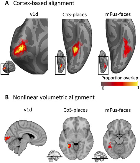
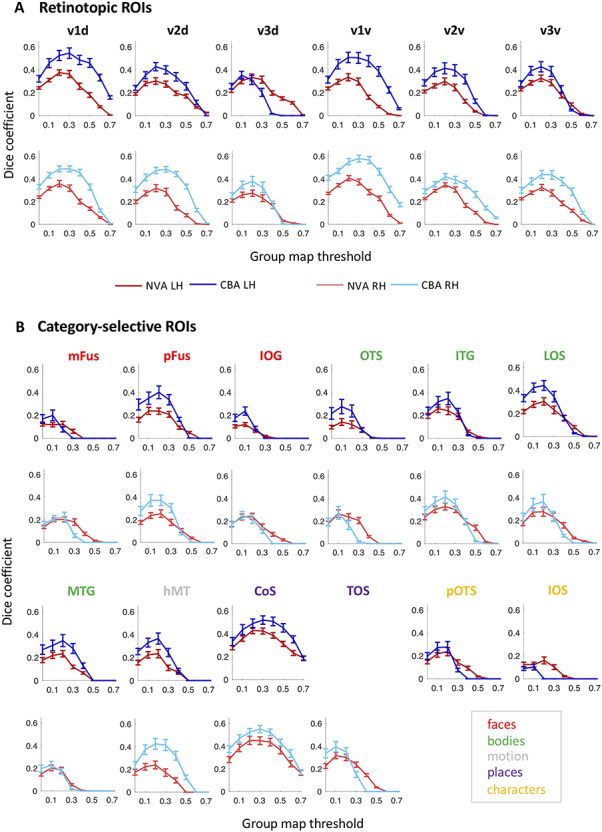
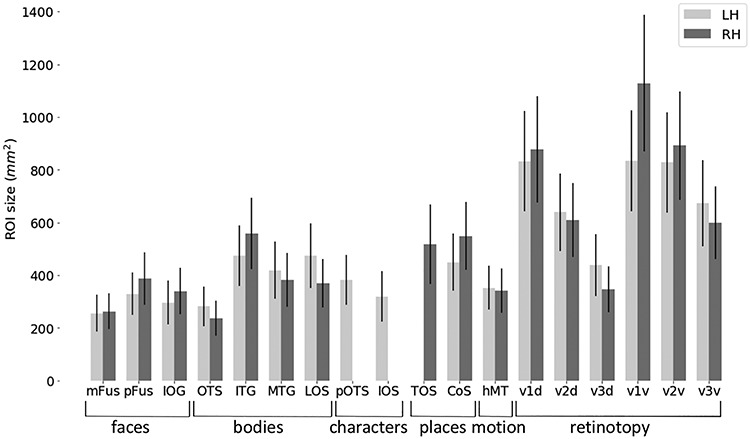
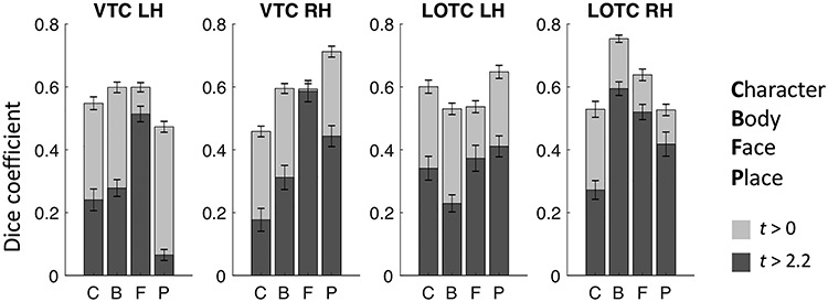
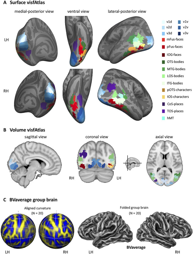
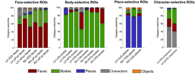
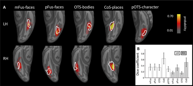
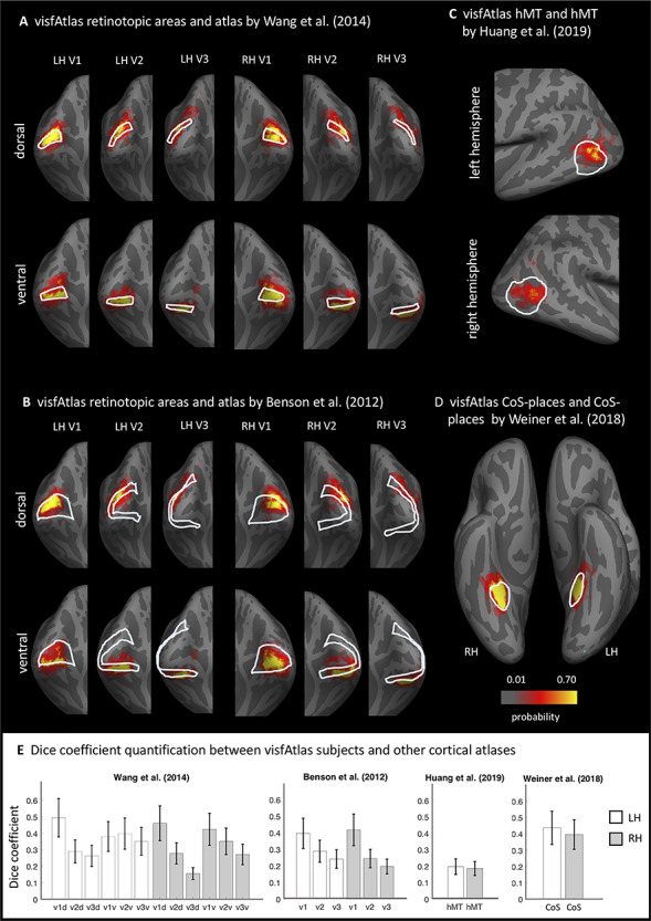
Human visual cortex contains many retinotopic and category-specific regions. These brain regions have been the focus of a large body of functional magnetic resonance imaging research, significantly expanding our understanding of visual processing. As studying these regions requires accurate localization of their cortical location, researchers perform functional localizer scans to identify these regions in each individual. However, it is not always possible to conduct these localizer scans. Here, we developed and validated a functional region of interest (ROI) atlas of early visual and category-selective regions in human ventral and lateral occipito-temporal cortex. Results show that for the majority of functionally defined ROIs, cortex-based alignment results in lower between-subject variability compared to nonlinear volumetric alignment. Furthermore, we demonstrate that 1) the atlas accurately predicts the location of an independent dataset of ventral temporal cortex ROIs and other atlases of place selectivity, motion selectivity, and retinotopy. Next, 2) we show that the majority of voxel within our atlas is responding mostly to the labeled category in a left-out subject cross-validation, demonstrating the utility of this atlas. The functional atlas is publicly available (download.brainvoyager.com/data/visfAtlas.zip) and can help identify the location of these regions in healthy subjects as well as populations (e.g., blind people, infants) in which functional localizers cannot be run.
Keywords: cortex-based alignment; human brain atlas; object recognition; retinotopy; visual cortex.
© The Author(s) 2020. Published by Oxford University Press.
Figures
Similar articles
-
A Retinotopic Basis for the Division of High-Level Scene Processing between Lateral and Ventral Human Occipitotemporal Cortex.J Neurosci. 2015 Aug 26;35(34):11921-35. doi: 10.1523/JNEUROSCI.0137-15.2015. J Neurosci. 2015. PMID: 26311774 Free PMC article. Clinical Trial.
-
Predicting Identity-Preserving Object Transformations across the Human Ventral Visual Stream.J Neurosci. 2021 Sep 1;41(35):7403-7419. doi: 10.1523/JNEUROSCI.2137-20.2021. Epub 2021 Jul 12. J Neurosci. 2021. PMID: 34253629 Free PMC article.
-
High-Level Representations in Human Occipito-Temporal Cortex Are Indexed by Distal Connectivity.J Neurosci. 2021 May 26;41(21):4678-4685. doi: 10.1523/JNEUROSCI.2857-20.2021. Epub 2021 Apr 13. J Neurosci. 2021. PMID: 33849949 Free PMC article.
-
Fine-scale spatial organization of face and object selectivity in the temporal lobe: do functional magnetic resonance imaging, optical imaging, and electrophysiology agree?J Neurosci. 2008 Nov 12;28(46):11796-801. doi: 10.1523/JNEUROSCI.3799-08.2008. J Neurosci. 2008. PMID: 19005042 Free PMC article. Review.
-
Human cortical areas underlying the perception of optic flow: brain imaging studies.Int Rev Neurobiol. 2000;44:269-92. doi: 10.1016/s0074-7742(08)60746-1. Int Rev Neurobiol. 2000. PMID: 10605650 Review.
Cited by
-
Natural scenes reveal diverse representations of 2D and 3D body pose in the human brain.Proc Natl Acad Sci U S A. 2024 Jun 11;121(24):e2317707121. doi: 10.1073/pnas.2317707121. Epub 2024 Jun 3. Proc Natl Acad Sci U S A. 2024. PMID: 38830105 Free PMC article.
-
Intracortical recordings reveal vision-to-action cortical gradients driving human exogenous attention.Nat Commun. 2024 Mar 26;15(1):2586. doi: 10.1038/s41467-024-46013-4. Nat Commun. 2024. PMID: 38531880 Free PMC article.
-
Both mOTS-words and pOTS-words prefer emoji stimuli over text stimuli during a reading task.bioRxiv [Preprint]. 2024 Jan 30:2023.11.07.565794. doi: 10.1101/2023.11.07.565794. bioRxiv. 2024. Update in: Cereb Cortex. 2024 Aug 1;34(8):bhae339. doi: 10.1093/cercor/bhae339. PMID: 37986766 Free PMC article. Updated. Preprint.
-
A personalized cortical atlas for functional regions of interest.J Neurophysiol. 2023 Nov 1;130(5):1067-1080. doi: 10.1152/jn.00108.2023. Epub 2023 Sep 20. J Neurophysiol. 2023. PMID: 37727907 Free PMC article.
-
Demystifying visual word form area visual and nonvisual response properties with precision fMRI.iScience. 2024 Nov 26;27(12):111481. doi: 10.1016/j.isci.2024.111481. eCollection 2024 Dec 20. iScience. 2024. PMID: 39759006 Free PMC article.
References
-
- Aguirre GK, Zarahn E, D’Esposito M. 1998. An area within human ventral cortex sensitive to “building” stimuli. Neuron. 21:373–383. - PubMed
-
- Amunts K, Malikovic A, Mohlberg H, Schormann T, Zilles K. 2000. Brodmann’s areas 17 and 18 brought into stereotaxic pace - where and how variable? Neuroimage. 11:66–84. - PubMed
-
- Barton JJS. 2008. Structure and function in acquired prosopagnosia: lessons from a series of 10 patients with brain damage. J Neuropsychol. 2:197. - PubMed
-
- Beauchamp MS, Haxby JV, Jennings JE, Deyoe EA. 1999. An fMRI version of the Farnsworth–Munsell 100-hue test reveals multiple color-selective areas in human ventral occipitotemporal cortex. Cereb Cortex. 9:257–263. - PubMed
Publication types
MeSH terms
Grants and funding
LinkOut - more resources
Full Text Sources
Research Materials
