Multicenter Study
. 2020 Oct 14;41(39):3827-3835.
doi: 10.1093/eurheartj/ehaa664.

Pathological features of COVID-19-associated myocardial injury: a multicentre cardiovascular pathology study

Affiliations
Multicenter Study

Pathological features of COVID-19-associated myocardial injury: a multicentre cardiovascular pathology study

Cristina Basso et al. Eur Heart J. .

Abstract

Aims: Coronavirus disease 2019 (COVID-19) due to severe acute respiratory syndrome coronavirus-2 (SARS-CoV-2) has been associated with cardiovascular features of myocardial involvement including elevated serum troponin levels and acute heart failure with reduced ejection fraction. The cardiac pathological changes in these patients with COVID-19 have yet to be well described.

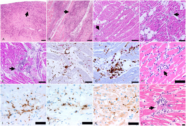
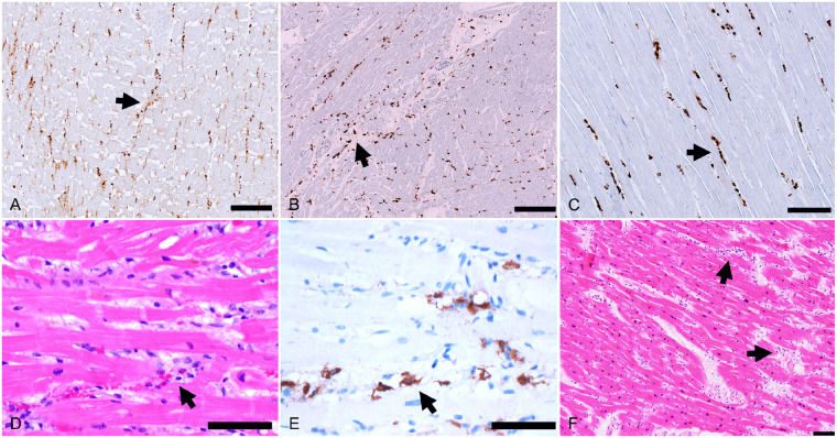
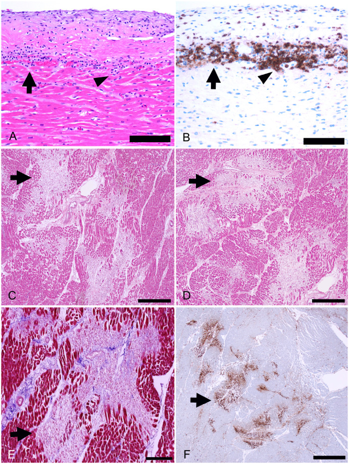
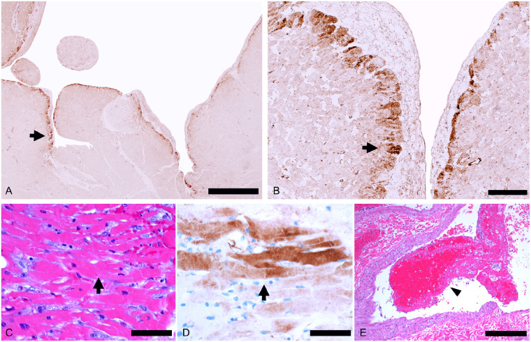
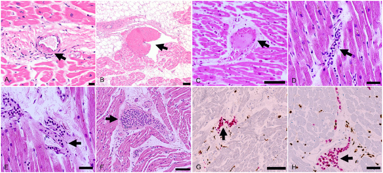
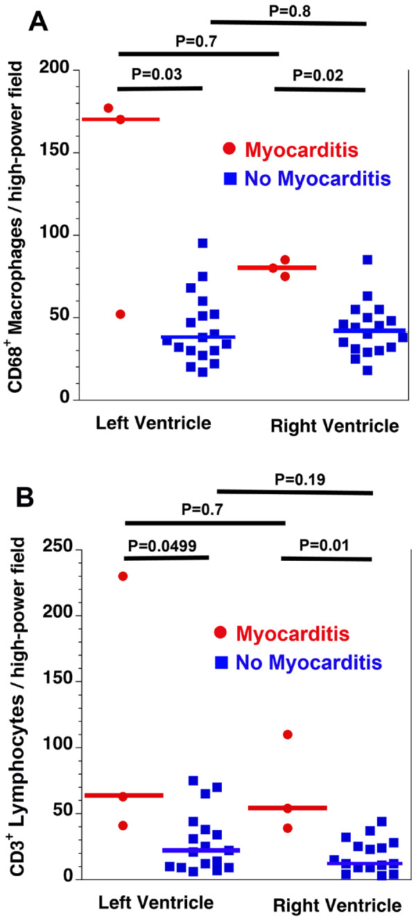
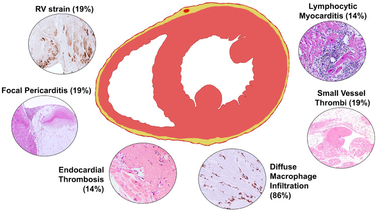
Methods and results: In an international multicentre study, cardiac tissue from the autopsies of 21 consecutive COVID-19 patients was assessed by cardiovascular pathologists. The presence of myocarditis, as defined by the presence of multiple foci of inflammation with associated myocyte injury, was determined, and the inflammatory cell composition analysed by immunohistochemistry. Other forms of acute myocyte injury and inflammation were also described, as well as coronary artery, endocardium, and pericardium involvement. Lymphocytic myocarditis was present in 3 (14%) of the cases. In two of these cases, the T lymphocytes were CD4 predominant and in one case the T lymphocytes were CD8 predominant. Increased interstitial macrophage infiltration was present in 18 (86%) of the cases. A mild pericarditis was present in four cases. Acute myocyte injury in the right ventricle, most probably due to strain/overload, was present in four cases. There was a non-significant trend toward higher serum troponin levels in the patients with myocarditis compared with those without myocarditis. Disrupted coronary artery plaques, coronary artery aneurysms, and large pulmonary emboli were not identified.

Conclusions: In SARS-CoV-2 there are increased interstitial macrophages in a majority of the cases and multifocal lymphocytic myocarditis in a small fraction of the cases. Other forms of myocardial injury are also present in these patients. The macrophage infiltration may reflect underlying diseases rather than COVID-19.

Keywords: Autopsy; COVID-19; Heart; Macrophages; Myocarditis; Myocardium; SARS; SARS-CoV-2.


Figures

Figure 1
Figure 1
Spectrum of myocarditis in COVID-19 patients. (A and B) Biventricular multifocal/diffuse lymphocytic myocarditis (arrows) with extensive myocyte injury in an 86-year-old man with previously undiagnosed cardiac amyloidosis (H&E ×50). (C–G) Biventricular multifocal lymphocytic myocarditis (arrows) with myocyte injury in a 64-year-old man, who developed atrial fibrillation 2 days before death (C, H&E ×100; D, H&E ×200; E, H&E ×100; F, double immunostaining CD68 brown/CD3 red, ×200; G, double immunostaining CD4 brown/CD8 red, ×400). (H–K) Biventricular multifocal lymphocytic myocarditis (arrow) in a 59-year-old man (H, H&E ×400; I, CD3 immunostaining brown, ×400; J, CD68 immunostaining brown, ×400; K, CD4 immunostaining brown, ×400). (L) Focal myocardial lymphocytic infiltration with myocyte injury (arrow) in a 70-year-old man (H&E ×400). Scale bars represent 500 μm (A, B), 200 μm (C, E), 100 μm (D, F), 50 μm (G–K), and 20 μm (L).
Figure 2
Figure 2
Increased interstitial macrophages. The majority of patients showed increased interstitial macrophages without associated myocyte injury (arrows). (A) A 50-year-old man, (CD68 immunostaining, ×100). (B) A 44-year-old man, (double CD68 brown/CD3 red immunostaining, ×100). (C) A 64-year-old man (CD68 immunostaining, ×200). (D and E) A 60-year-old man (D, H&E, ×400; (E) CD68 immunostaining, ×400). (F) A 73-year-old woman with increased cells within the myocardial interstitium (H&E, ×100). Scale bars represent 200 μm (A, B), 100 μm (C, F), and 50 μm (D, E).
Figure 3
Figure 3
Other myocardial inflammatory changes in patients with COVID-19. (A and B) Focal lymphocytic pericarditis (arrows), mainly composed of CD8+ lymphocytes, associated with focal myocardial inflammation without myocyte injury (arrowhead) in a 66-year-old man (A, H&E, ×400; B, CD8 immunostaining, ×400). (C–F) Healing myocardial injury (arrows) in the subendocardium of the left ventricle in a 50-year-old man (C and D, H&E, ×50; E, Azan Mallory trichrome, ×100; F, CD68 immunostaining, ×25). Scale bars represent 50 μm (A, B), 500 μm (C, D), 200 μm (E) and 1000 μm (F).
Figure 4
Figure 4
Right ventricle myocardial injury consistent with strain. (A and B) Right ventricle myocardium showing subendocardial C4d+ necrotic myocytes (arrows) in a 50-year-old man (C4d immunostaining: A ×25; B ×200). (C–E) C4d + necrotic myocytes (arrows) in the right ventricle in a 60-year-old woman with microvascular pulmonary arterial thrombi (E, arrowhead) (C, H&E, ×400; D, C4d immunostaining 4 × 00; E, H&E ×100). Scale bars represent 1000 μm (A), 100 μm (B), 50 μm (C, D), and 200 μm (E).
Figure 5
Figure 5
Myocardial small vessel changes. (A and B) Organizing microthrombus (arrow) in a small myocardial artery (A, H&E, ×400) and organizing venous thrombus (arrow) (B, H&E, ×100) in a 70-year-old man. (C) Thrombus (arrow) in a small myocardial vein (H&E, ×200) in a 71-year-old man. (D and E) Leucocyte aggregates (mainly neutrophils and mononuclear cells, arrows) in capillaries and small veins in a 64-year-old man (H&E, ×400). (F–H) A 44-year-old man with eosinophils and mononuclear cells (arrow) filling the lumen of a small dilated vein (F, H&E, ×200) and intravascular T lymphocytes (arrows) in myocardial small veins (double immunostaning CD68 brown/CD3 red: G ×200; H ×400). Scale bars represent 20 μm (A), 100 μm (B, C, F, G), and 50 μm (D, E, H).
Figure 6
Figure 6
Quantification of inflammatory cells. There were more CD68+ macrophages (A) and CD3+ lymphocytes (B) in the myocarditis group compared with the no myocarditis group. There were no differences in inflammatory cell density between the left and right ventricles for either group. Horizontal bars indicate the median values. Overall P = 0.01 (A) and P = 0.005 (B).
Figure 7
Figure 7
Cardiac pathological changes associated with COVID-19.

Comment in

References

    1. Zhu N, Zhang D, Wang W, Li X, Yang B, Song J, Zhao X, Huang B, Shi W, Lu R, Niu P, Zhan F, Ma X, Wang D, Xu W, Wu G, Gao GF, Tan W. A novel coronavirus from patients with pneumonia in China 2019. N Engl J Med 2020;382:727–733. - PMC - PubMed
    1. Shi S, Qin M, Shen B, Cai Y, Liu T, Yang F, Gong W, Liu X, Liang J, Zhao Q, Huang H, Yang B, Huang C. Association of cardiac injury with mortality in hospitalized patients with COVID-19 in Wuhan, China. JAMA Cardiol 2020;5:802–210. - PMC - PubMed
    1. Zhou F, Yu T, Du R, Fan G, Liu Y, Liu Z, Xiang J, Wang Y, Song B, Gu X, Guan L, Wei Y, Li H, Wu H, Xu J, Tu S, Zhang Y, Chen H, Cao B. Clinical course and risk factors for mortality of adult inpatients with COVID-19 in Wuhan, China: a retrospective cohort study. Lancet 2020;395:1054–1062. - PMC - PubMed
    1. Lippi G, Lavie CJ, Sanchis-Gomar F. Cardiac troponin I in patients with coronavirus disease 2019 (COVID-19): evidence from a meta-analysis. Prog Cardiovasc Dis 2020;doi: 10.1016/j.pcad.2020.03.001. - PMC - PubMed
    1. Inciardi RM, Lupi L, Zaccone G, Italia L, Raffo M, Tomasoni D, Cani DS, Cerini M, Farina D, Gavazzi E, Maroldi R, Adamo M, Ammirati E, Sinagra G, Lombardi CM, Metra M. Cardiac involvement in a patient with coronavirus disease 2019 (COVID-19). JAMA Cardiol 2020;5:819–824. - PMC - PubMed

Publication types