. 2020 Nov 17;53(11):2600-2610.
doi: 10.1021/acs.accounts.0c00434. Epub 2020 Sep 24.

Molecular Photoswitching in Confined Spaces

Affiliations

Molecular Photoswitching in Confined Spaces

Angela B Grommet et al. Acc Chem Res. .

Abstract

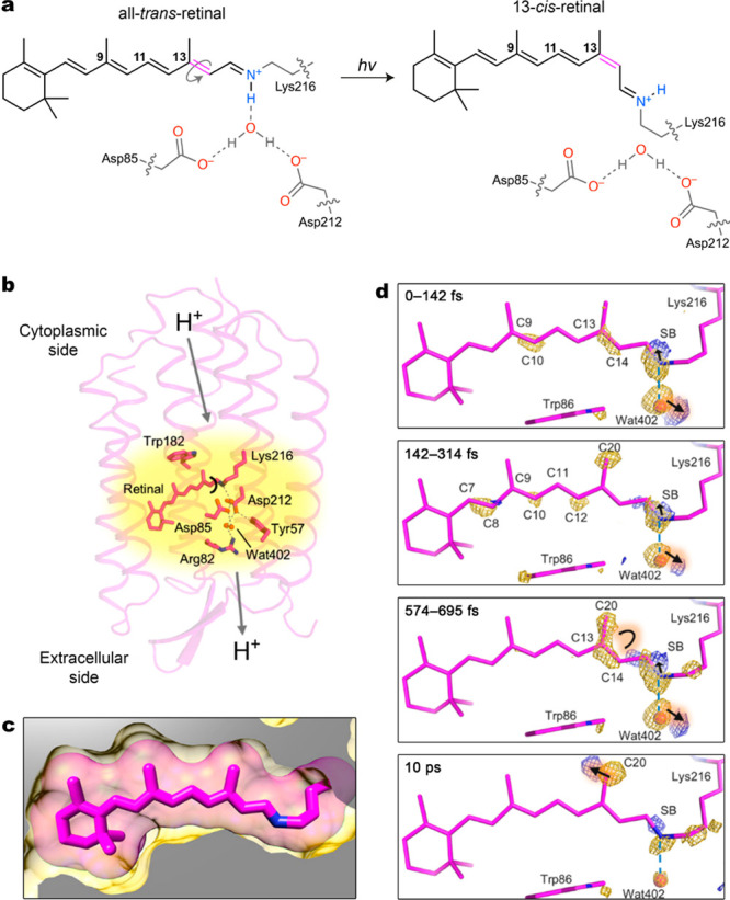
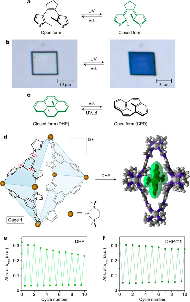
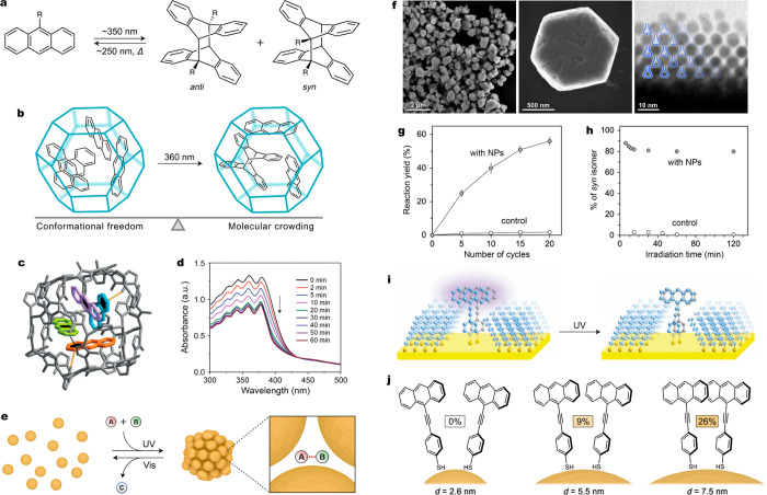
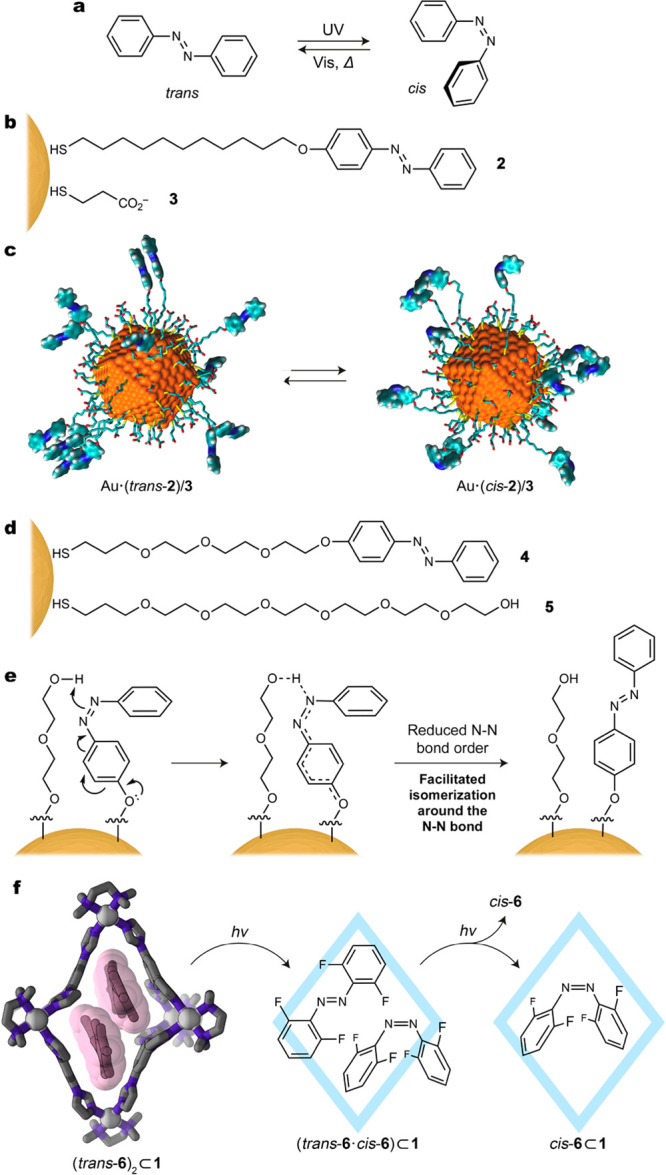
In nature, light is harvested by photoactive proteins to drive a range of biological processes, including photosynthesis, phototaxis, vision, and ultimately life. Bacteriorhodopsin, for example, is a protein embedded within archaeal cell membranes that binds the chromophore retinal within its hydrophobic pocket. Exposure to light triggers regioselective photoisomerization of the confined retinal, which in turn initiates a cascade of conformational changes within the protein, triggering proton flux against the concentration gradient, providing the microorganisms with the energy to live. We are inspired by these functions in nature to harness light energy using synthetic photoswitches under confinement. Like retinal, synthetic photoswitches require some degree of conformational flexibility to isomerize. In nature, the conformational change associated with retinal isomerization is accommodated by the structural flexibility of the opsin host, yet it results in steric communication between the chromophore and the protein. Similarly, we strive to design systems wherein isomerization of confined photoswitches results in steric communication between a photoswitch and its confining environment. To achieve this aim, a balance must be struck between molecular crowding and conformational freedom under confinement: too much crowding prevents switching, whereas too much freedom resembles switching of isolated molecules in solution, preventing communication.In this Account, we discuss five classes of synthetic light-switchable compounds-diarylethenes, anthracenes, azobenzenes, spiropyrans, and donor-acceptor Stenhouse adducts-comparing their behaviors under confinement and in solution. The environments employed to confine these photoswitches are diverse, ranging from planar surfaces to nanosized cavities within coordination cages, nanoporous frameworks, and nanoparticle aggregates. The trends that emerge are primarily dependent on the nature of the photoswitch and not on the material used for confinement. In general, we find that photoswitches requiring less conformational freedom for switching are, as expected, more straightforward to isomerize reversibly under confinement. Because these compounds undergo only small structural changes upon isomerization, however, switching does not propagate into communication with their environment. Conversely, photoswitches that require more conformational freedom are more challenging to switch under confinement but also can influence system-wide behavior.Although we are primarily interested in the effects of geometric constraints on photoswitching under confinement, additional effects inevitably emerge when a compound is removed from solution and placed within a new, more crowded environment. For instance, we have found that compounds that convert to zwitterionic isomers upon light irradiation often experience stabilization of these forms under confinement. This effect results from the mutual stabilization of zwitterions that are brought into close proximity on surfaces or within cavities. Furthermore, photoswitches can experience preorganization under confinement, influencing the selectivity and efficiency of their photoreactions. Because intermolecular interactions arising from confinement cannot be considered independently from the effects of geometric constraints, we describe all confinement effects concurrently throughout this Account.


Conflict of interest statement

The authors declare no competing financial interest.

Figures

Figure 1
Figure 1
(a) Light-induced regioselective isomerization of retinal within the binding pocket of bacteriorhodopsin. (b) Structure of bacteriorhodopsin with retinal buried inside its hydrophobic cavity. The arrows indicate the direction of light-induced proton transfer. (c) Binding of retinal (pink sticks with van der Waals radii shown as a transparent halo) encased within the hydrophobic cavity of bacteriorhodopsin (yellow). (d) Structural dynamics of retinal and its immediate surroundings captured by a femtosecond X-ray laser. The transition from trans-retinal to cis-retinal is mapped onto a dark-state model based on the difference Fourier electron density (FobslightFobsdark) contoured at 4σ (yellow, negative; blue, positive). Adapted with permission from ref (5). Copyright 2018 American Association for the Advancement of Science.
Figure 2
Figure 2
(a) Reversible photoisomerization of a diarylethene. The colored isomer featuring extensive conjugation of π electrons (here, the closed form) is shown in green. (b) Reversible light-induced deformation of a single crystal of a simple diarylethene (here, 1,2-bis(2-ethyl-5-phenyl-3-thienyl)perfluorocyclopentene). (c) Reversible photoisomerization between dihydropyrene (DHP) and cyclophanediene (CPD). (d) Structural formula of coordination cage 1 used to investigate the behavior of photoswitchable molecules under confinement (left) and crystal structure of an inclusion complex of DHP inside cage 1 (right). (e) Gradual decomposition of DHP in pentane solution over 10 switching cycles. (f) Improved fatigue resistance of DHP⊂1 over 10 cycles under the same irradiation conditions. (b) Adapted with permission from ref (24). Copyright 2007 Springer Nature. (e, f) Adapted from ref (31). Copyright 2020 American Chemical Society.
Figure 3
Figure 3
(a) Reversible photodimerization of anthracene. (b) Proposed photoreaction of anthracene inside the cavity of ZIF-8. (c) Crystal structure of a ZIF-8 cavity encapsulating four molecules of anthracene. (d) UV/vis absorption spectra accompanying UV irradiation of ZIF-8 encapsulating anthracene. (e) Schematic illustration of light-induced trapping and increased reactivity of small molecules within colloidal crystals (“dynamically self-assembling nanoflasks”). (f) Electron micrographs (at different magnifications) of colloidal crystals prepared by exposing azobenzene-coated gold nanoparticles to UV light. (g) Accelerated photodimerization of 9-anthracenemethanol in the presence of photoresponsive nanoparticles. (h) Stereoselectivity in the dimerization of 9-anthracenemethanol in the presence and absence of photoresponsive nanoparticles. (i) Schematic illustration of the photoreaction of 9-(4-mercaptophenylethynyl)anthracene on a Au(111) surface. (j) Dependence of the anthracene dimerization yield on the curvature of the underlying nanoparticle. (c, d) Adapted with permission from ref (34). Copyright 2019 Wiley-VCH. (f–h) Adapted with permission from ref (4). Copyright 2016 Springer Nature. (i) Adapted with permission from ref (37). Copyright 2011 American Association for the Advancement of Science.
Figure 4
Figure 4
(a) Reversible photoisomerization of azobenzene. (b) Structural formulas of azobenzene 2 and background ligand 3. (c) Snapshots from molecular dynamics simulations of a 2/3-coated gold nanoparticle in the trans and cis states of azobenzene. (d) Structural formulas of azobenzene 4 and background ligand 5. (e) Proposed mechanism for accelerated azobenzene isomerization on a nanoparticle surface upon coadsorption with a hydroxy-terminated background ligand. (f) Crystal structure of an inclusion complex comprising cage 1 and two molecules of tetra-o-fluoroazobenzene (6) (left) and the stepwise mechanism underlying the photoisomerization of 6 within the cavity of 1 (right). (c) Adapted from ref (3). Copyright 2019 American Chemical Society.
Figure 5
Figure 5
(a, b) Reversible photoisomerization of (a) spiropyran and (b) DASA. (c) Comparison of the kinetics of merocyanine→spiropyran back-isomerization in solution and on the surface of 2.6 nm gold nanoparticles (χ = 0.33). (d) Dependence of the kinetics of disassembly of gold nanoparticle aggregates on the spiropyran coverage. (e, f) Spontaneous bleaching of the open form of DASA on the surfaces of (e) 4.2 and (f) 8.6 nm magnetite nanoparticles. (g) TEM image of a spiropyran-incorporating framework. The arrows indicate individual nanopores. (h) Encapsulation of spiropyran 7 within cage 1 and the resulting crystal structure (hydrogen atoms of the cage have been omitted for clarity). (i) Mechanism of the light-induced decoloration of 71. (c, d) From ref (55). CC BY 3.0. (e, f) Adapted with permission from ref (57). Copyright 2017 Wiley-VCH. (g) From ref (2). CC BY NC ND 3.0.

References

    1. Samanta D.; Gemen J.; Chu Z.; Diskin-Posner Y.; Shimon L. J. W.; Klajn R. Reversible photoswitching of encapsulated azobenzenes in water. Proc. Natl. Acad. Sci. U. S. A. 2018, 115, 9379–9384. 10.1073/pnas.1712787115. - DOI - PMC - PubMed
    1. Kundu P. K.; Olsen G. L.; Kiss V.; Klajn R. Nanoporous frameworks exhibiting multiple stimuli responsiveness. Nat. Commun. 2014, 5, 3588.10.1038/ncomms4588. - DOI - PubMed
    1. Chu Z.; Han Y.; Bian T.; De S.; Král P.; Klajn R. Supramolecular Control of Azobenzene Switching on Nanoparticles. J. Am. Chem. Soc. 2019, 141, 1949–1960. 10.1021/jacs.8b09638. - DOI - PubMed
    1. Zhao H.; Sen S.; Udayabhaskararao T.; Sawczyk M.; Kučanda K.; Manna D.; Kundu P. K.; Lee J.-W.; Král P.; Klajn R. Reversible trapping and reaction acceleration within dynamically self-assembling nanoflasks. Nat. Nanotechnol. 2016, 11, 82–88. 10.1038/nnano.2015.256. - DOI - PubMed
    1. Nogly P.; Weinert T.; James D.; Carbajo S.; Ozerov D.; Furrer A.; Gashi D.; Borin V.; Skopintsev P.; Jaeger K.; Nass K.; Båth P.; Bosman R.; Koglin J.; Seaberg M.; Lane T.; Kekilli D.; Brünle S.; Tanaka T.; Wu W.; Milne C.; White T.; Barty A.; Weierstall U.; Panneels V.; Nango E.; Iwata S.; Hunter M.; Schapiro I.; Schertler G.; Neutze R.; Standfuss J. Retinal isomerization in bacteriorhodopsin captured by a femtosecond X-ray laser. Science 2018, 361, eaat0094.10.1126/science.aat0094. - DOI - PubMed

Publication types