

PARACENTRAL ACUTE MIDDLE MACULOPATHY ASSOCIATED WITH BRANCH RETINAL ARTERY OCCLUSION DUE TO POLYCYTHEMIA IN A PATIENT WITH TETRALOGY OF FALLOT
- PMID: 32969982
- PMCID: PMC9394495
- DOI: 10.1097/ICB.0000000000001054
PARACENTRAL ACUTE MIDDLE MACULOPATHY ASSOCIATED WITH BRANCH RETINAL ARTERY OCCLUSION DUE TO POLYCYTHEMIA IN A PATIENT WITH TETRALOGY OF FALLOT
Abstract
Purpose: To describe the occurrence of paracentral acute middle maculopathy (PAMM) associated with branch retinal artery occlusion secondary to polycythemia in a patient with tetralogy of Fallot.
Methods: Case report.
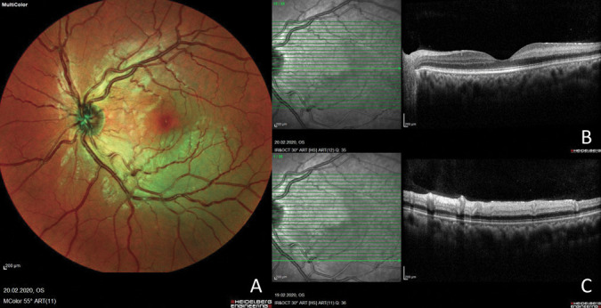
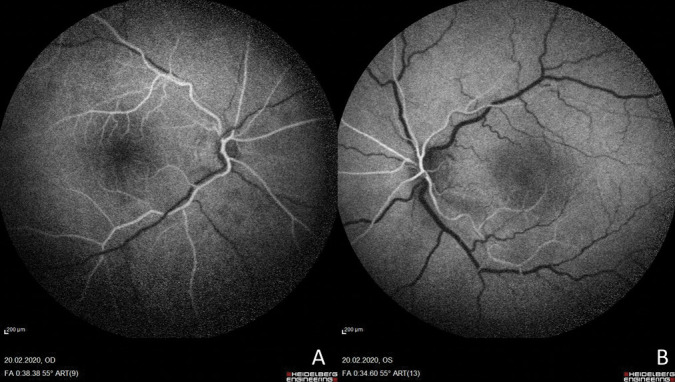
Results: A 30-year-old man presented with acute vision loss and superior visual deficit in his left eye for two days. His medical record had a tetralogy of Fallot. Complete blood count showed an erythrocyte count of 9.88 million/µL (4.4-5.6), hemoglobin of 17.7 g/dL (13.5-16.9), and hematocrit of 65.4% (40-49). The best-corrected visual acuity was 20/25 in the left eye, and a diagnosis of left inferotemporal branch retinal artery occlusion was made. Spectral-domain optical coherence tomography revealed a characteristic hyperreflective band-like lesion on the inner nuclear layer consistent with PAMM.
Conlusion: Polycythemia may be a trigger for branch retinal artery occlusion-associated PAMM. We suggest a new precursor cause of PAMM that is previously undescribed.
Copyright © 2020 The Author(s). Published by Wolters Kluwer Health, Inc. on behalf of the Opthalmic Communications Society, Inc.
Conflict of interest statement
None of the authors has any financial/conflicting interests to disclose.
Figures
References
-
- Sarraf D, Rahimy E, Fawzi AA, et al. Paracentral acute middle maculopathy: a new variant of acute macular neuroretinopathy associated with retinal capillary ischemia. JAMA Ophthalmol 2013;131:1275–1287. - PubMed
-
- Rahimy E, Sarraf D, Dollin ML, et al. Paracentral acute middle maculopathy in nonischemic central retinal vein occlusion. Am J Ophthalmol 2014;158:372–380.e1. - PubMed
-
- Chen X, Rahimy E, Sergott RC, et al. Spectrum of retinal vascular diseases associated with paracentral acute middle maculopathy. Am J Ophthalmol 2015;160:26–34.e1. - PubMed
-
- Shahlaee A, Sridhar J, Rahimy E, et al. Paracentral acute middle maculopathy associated with postviral Purtscher-like retinopathy. Retin Cases Brief Rep 2019;13:50–53. - PubMed
-
- Ilginis T, Keane PA, Tufail A. Paracentral acute middle maculopathy in sickle cell disease. JAMA Ophthalmol 2015;133:614–616. - PubMed
Publication types
MeSH terms
LinkOut - more resources
Full Text Sources
Medical
