Review
. 2020 Nov;125(11):1072-1086.
doi: 10.1007/s11547-020-01287-8. Epub 2020 Sep 24.

Cardiovascular magnetic resonance (CMR) in restrictive cardiomyopathies

Affiliations
Review

Cardiovascular magnetic resonance (CMR) in restrictive cardiomyopathies

Nicola Galea et al. Radiol Med. 2020 Nov.

Abstract

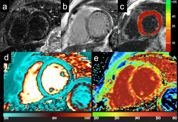
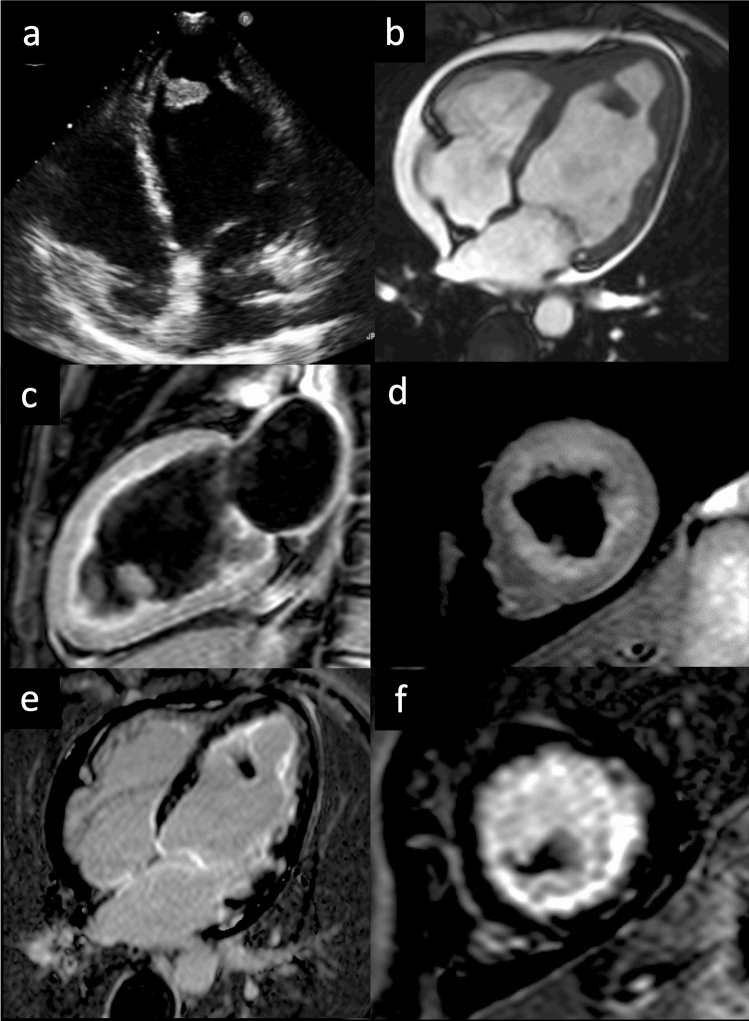
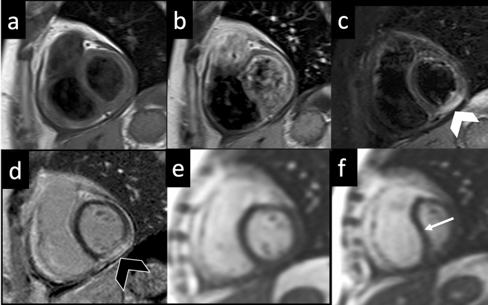
The restrictive cardiomyopathies constitute a heterogeneous group of myocardial diseases with a different pathogenesis and overlapping clinical presentations. Diagnosing them frequently poses a challenge. Echocardiography, electrocardiograms and laboratory tests may show non-specific changes. In this context, cardiac magnetic resonance (CMR) may play a crucial role in defining the diagnosis and guiding treatments, by offering a robust myocardial characterization based on the inherent magnetic properties of abnormal tissues, thus limiting the use of endomyocardial biopsy. In this review article, we explore the role of CMR in the assessment of a wide range of myocardial diseases causing restrictive patterns, from iron overload to cardiac amyloidosis, endomyocardial fibrosis or radiation-induced heart disease. Here, we emphasize the incremental value of novel relaxometric techniques such as T1 and T2 mapping, which may recognize different storage diseases based on the intrinsic magnetic properties of the accumulating metabolites, with or without the use of gadolinium-based contrast agents. We illustrate the importance of these CMR techniques and their great support when contrast media administration is contraindicated. Finally, we describe the useful role of cardiac computed tomography for diagnosis and management of restrictive cardiomyopathies when CMR is contraindicated.

Keywords: Cardiac imaging; Cardiovascular magnetic resonance; Infiltrative cardiomyopathies; Restrictive cardiomyopathies.


Conflict of interest statement

The authors declare that they have no conflict of interest.

Figures

Fig. 1
Fig. 1
57-year-old patient with multiple myeloma with known bone lesion associated with light chain proteinuria and bilateral carpal tunnel syndrome. Cine-SSFP sequences (a short axis view; b 4-chamber view) showed a thickening of the left ventricular myocardium wall (19 mm in the septum) with global and moderate hypokinesia (left ventricular ejection fraction 46%). In panel c are reported some of the images of the lock–locker sequences of the TI scout. Inversion recovery turbo field echo sequences (d and e short axis view; f and g 4-chamber view) with a wrong myocardium null time (red box and arrow) and the one with the correct null time (green box and arrow); these last showed a diffuse areas of circumferential subendocardial pattern enhancement. The final diagnosis was light chain (AL) cardiac amyloidosis
Fig. 2
Fig. 2
83-year-old male patient with known CAD and the presence of dyspnoea. Cine-SSFP sequences (a short axis view; b 4-chamber view), which show a thickening of both the left ventricular myocardium (18 mm in the septum) and the right ventricle, but also of the atrial walls with global and severe hypokinesia (left ventricular ejection fraction 26%). Inversion recovery turbo field echo sequences (c short axis view; d 4-chamber view) for late gadolinium enhancement (LGE) analysis; there are diffuse areas of circumferential subendocardial pattern enhancement even with transmural extension in the basal segment. There is also LGE within the right ventricle and both atrial walls. The quantitative evaluation of global left ventricular myocardium native T1 (e short axis view) and ECV (f short axis view) resulted in 1110 ms (v.n. 1000 ms) and 55% (v.n. 20–30%), respectively. Overall, the presence and the pattern of LGE with a transmural pattern in both ventricle and atrial walls were suspicious of transthyretin (ATTR) amyloidosis. The patient was then scanned with 99mTc-DPD (image g), where the abnormal and diffuse presence of the osteotropic indicator is observed in the left and right ventricle with a Perugini score = 3. The final diagnosis was ATTR amyloidosis
Fig. 3
Fig. 3
57-year-old female with frequent syncopal episodes and ventricular tachycardia, LV dilation and severe reduction in EF at TTE, with no obstruction of coronary arteries at coronary angiography. CMR revealed no edema on T2w-STIR images (a short axis view and d LV long axis view) and extensive areas of late gadolinium enhancement at IR-TFE images (b short axis view and e LV long axis view), with a non-ischemic pattern of distribution. The FDG-PET (c short axis view, f long axis view) confirmed the diagnosis of sarcoidosis with the identification of areas of FDG uptake (i.e., active inflammation) within the myocardium and in the mediastinal lymph nodes. LV left ventricle, EF ejection fraction; TTE transthoracic echocardium
Fig. 4
Fig. 4
Anderson–Fabry disease—Cine-SSFP in short axis (a) and four-chamber (b) views acquired on end-diastolic phase demonstrate an asymmetrical hypertrophy with predominant involvement of septum (IVS maximal thickness: 20 mm). On LGE image (c), an area of mid-myocardial enhancement is detected in the LV inferolateral wall (red arrow). STIR T2-weighted image (d) shows an area of myocardial edema located in LV antero-lateral wall, with a subendocardial distribution pattern (white arrow), confirmed by the blue area (T2 ratio > 2) in the panel at the bottom. The analysis of nT1 (e) map demonstrates severe reduction in global nT1 (reddish brown color, nT1: 877 ± 23 ms, normal value for our scanner 970–1020 ms) except for the focus of increased nT1 at inferolateral wall (nT1: 1116 ms, black arrowhead) matching the area of increase in ECV (white arrowhead, ECV: 48%) on relative map (f, global ECV: 27%). Hematoxylin and eosin histology (g, ×200) shows cardiomyocytes hypertrophy, caused by large cytoplasmic and perinuclear vacuoles, containing myelin bodies. Cine-SSFP steady-state free precession images; IVS interventricular septum; LGE late gadolinium enhancement; STIR short tau inversion recovery; LV left ventricle; nT1 native T1 map; ECV extracellular volume fraction
Fig. 5
Fig. 5
Cardiac iron overload—A 42-year-old woman with Cooley’s disease and moderate reduction in ventricular function (EF: 42%) show a global myocardial hypointensity on STIR image (a) and a diffuse inhomogeneous abnormal signal on LGE imaging (b), with no evidence of clear focal areas of enhancement. Analysis of T2* map (c), generated by traditional multiecho gradient echo T2-weighted sequence, shows a diffuse and marked reduction in the global myocardial T2* relaxation time (T2* = 0–1.5 ms, normal value > 20 ms). nT1 map (d) shows a significant reduction in global nT1 value (nT1 ≈ 535 ms, normal value 970–1020 ms), affected by susceptibility effect of intramyocardial iron accumulation. ECV map e reveals diffuse fibrosis (ECV ≈ 38–40%). STIR short tau inversion recovery; LGE late gadolinium enhancement; nT1 native T1 value; ECV extracellular volume fraction
Fig. 6
Fig. 6
Endomyocardial fibrosis—40-year-old patient with hypereosinophilia. Transthoracic echocardiogram in 4-chamber projection demonstrates the presence of an apical thrombus (a). Cine-SSFP in 4-chamber view (b) confirms echocardiographic findings and evidence a reduction in EF (35%). STIR in 2-chamber plane (c) and short axis (d) shows no signs of edema. LGE images in 4 chambers (e) and short axis (f) demonstrate a diffuse subendocardial hyperintensity and a circumferential pericardial effusion
Fig. 7
Fig. 7
Constrictive pericarditis—TSE T1-weighted images acquired on short axis view before (a) and 3 min after gadolinium administration (b) demonstrate a diffuse thickening of pericardial layer with minimal effusion and slight enhancement of the visceral pericardial layer. On STIR T2-weighted image (c), a focal area on myocardial edema (white arrowhead) is found in the LV inferior wall corresponding to the area of pathological enhancement (black arrowhead) on LGE image (d) by demonstrating a condition of active myocarditis. On free-breathing real-time cine-SSFP acquired in end-expiration (e) and end-inspiration (f), a bouncing and leftward shifting of IVS is seen during inspiration (white arrow) due to the inversion of interventricular pressure ratio combined with inextensibility of the pericardial sac. TSE turbo spin echo; STIR short tau inversion recovery; LGE late gadolinium enhancement; LV left ventricle; SSFP steady-state free precession; IVS interventricular septum

References

    1. Pereira NL, Grogan M, Dec GW. Spectrum of restrictive and infiltrative cardiomyopathies: part 1 of a 2-part series. J Am Coll Cardiol. 2018;71:1130–1148. doi: 10.1016/j.jacc.2018.01.016. - DOI - PubMed
    1. White SK, Sado DM, Fontana M, et al. T1 mapping for myocardial extracellular volume measurement by CMR: bolus only versus primed infusion technique. JACC Cardiovasc Imaging. 2013;6:955–962. doi: 10.1016/j.jcmg.2013.01.011. - DOI - PubMed
    1. Muehlberg F, Toepper A, Fritschi S, et al. Magnetic resonance imaging applications on infiltrative cardiomyopathies. J Thorac Imaging. 2016;31:336–347. doi: 10.1097/RTI.0000000000000199. - DOI - PubMed
    1. Dorbala S, Cuddy S, Falk RH. How to image cardiac amyloidosis: a practical approach. JACC Cardiovasc Imaging. 2019 doi: 10.1016/j.jcmg.2019.07.015. - DOI - PMC - PubMed
    1. Fontana M, Ćorović A, Scully P, Moon JC. Myocardial amyloidosis: the exemplar interstitial disease. JACC Cardiovasc Imaging. 2019;12:2345–2356. doi: 10.1016/j.jcmg.2019.06.023. - DOI - PubMed

MeSH terms

LinkOut - more resources