Review
. 2021 May;157(4):930-943.
doi: 10.1111/jnc.15197. Epub 2020 Oct 12.

Viral infection and smell loss: The case of COVID-19

Affiliations
Review

Viral infection and smell loss: The case of COVID-19

Isaias Glezer et al. J Neurochem. 2021 May.

Abstract

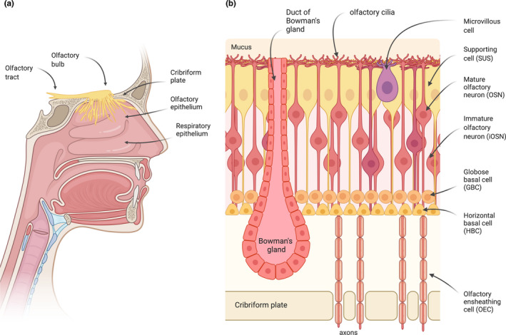
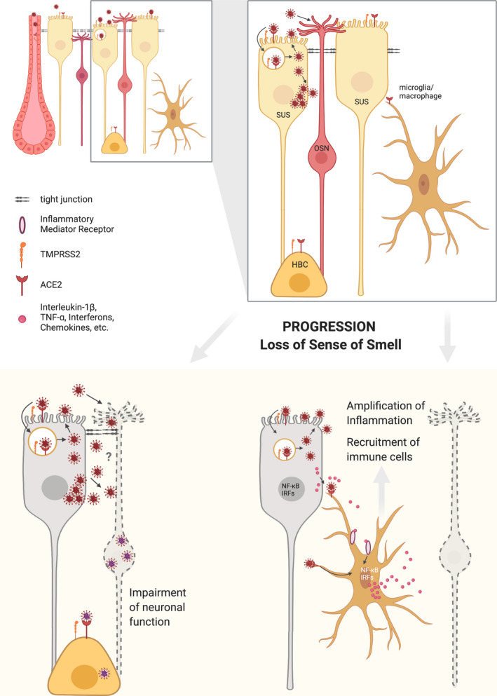
Olfactory disorders have been increasingly reported in individuals infected with SARS-CoV-2, the virus causing the coronavirus disease 2019 (COVID-19). Losing the sense of smell has a strong impact on the quality of life, since it may lead to malnutrition, weight loss, food poisoning, depression, and exposure to dangerous chemicals. Individuals who suffer from anosmia (inability to smell) also cannot sense the flavor of food, which is a combination of taste and smell. Interestingly, infected individuals have reported sudden loss of smell with no congested nose, as is frequently observed in common colds or other upper respiratory tract infections. These observations suggest that SARS-CoV-2 infection leads to olfactory loss through a distinct mechanism, which is still unclear. This article provides an overview of olfactory loss and the recent findings relating to COVID-19. Possible mechanisms of SARS-CoV-2-induced olfactory loss are also discussed.

Keywords: SARS-CoV-2; anosmia; coronavirus; olfaction; olfactory sensory neuron; smell loss.


Figures

FIGURE 1
FIGURE 1
The olfactory system. (a) Odorants are detected by OSNs present in the olfactory epithelium (OE), a specialized neuroepithelium located in the highest recesses of the nose. The olfactory sensory neurons (OSNs) project a single unmyelinated axon to the olfactory bulb (OB). The axons of the OSNs and their associated olfactory ensheathing cells (OECs) form bundles that project through the perforations of the cribriform plate to the OB, where they synapse with the mitral and tufted cells, forming the glomeruli. The axons of these cells form the olfactory tract, which transmits the sensory information into the brain. (b) The different cell types that compose the olfactory epithelium are represented. Odorants are recognized by odorant receptors located in the cilia of the OSNs.
FIGURE 2
FIGURE 2
SARS‐CoV‐2 and olfactory neuron function. Supporting cells (SUS), horizontal basal cells (HBC) and Bowmans' gland cells (but not OSNs) express the SARS‐CoV‐2 entry factors ACE2 and TMPRSS2. It is therefore likely that SARS‐CoV‐2 primarily infects the supporting cells and gland cells which are located in the apical region of the epithelium. Damage to the supporting cells would indirectly lead to disruption of proper odorant signaling by OSNs. It remains to be determined whether the virus could infect the OSNs in an ACE2‐independent manner and directly interfere with their function. Importantly, infected supporting cells can release molecules that trigger innate immune signaling in resident microglia/macrophages, which are also responsive to the viral particles. In consequence, key pro‐inflammatory transcription factors (such as NF‐kappaB and IRFs) promote synthesis of interferons and inflammatory mediators that recruit and activate varied types of leukocytes. The olfactory neurons are vulnerable to inflammation, resulting in temporary olfactory loss until the viral infection is resolved and they are replenished by new neurons. Horizontal basal cells (HBCs), which are essential for regeneration of the epithelium after profound damage, express the SARS‐CoV‐2 entry proteins. Whether the virus can also infect these cells and perturb regeneration of the epithelium, remains unknown.

References

    1. Adney, D. R. , van Doremalen, N. , Brown, V. R. , Bushmaker, T. , Scott, D. , de Wit, E. , Bowen, R. A. , & Munster, V. J. (2014). Replication and shedding of MERS‐CoV in upper respiratory tract of inoculated dromedary camels. Emerging Infectious Diseases, 20, 1999–2005. 10.3201/eid2012.141280 - DOI - PMC - PubMed
    1. Allavena, R. E. , Desai, B. , Goodwin, D. , Khodai, T. , & Bright, H. (2011). Pathologic and virologic characterization of neuroinvasion by HSV‐2 in a mouse encephalitis model. Journal of Neuropathology and Experimental Neurology, 70, 724–734. 10.1097/NEN.0b013e3182275264 - DOI - PubMed
    1. Aragao, M. , Leal, M. C. , Cartaxo Filho, O. Q. , Fonseca, T. M. , & Valenca, M. M. (2020). Anosmia in COVID‐19 associated with injury to the olfactory bulbs evident on MRI. AJNR. American Journal of Neuroradiology, 41, 1703–1706. 10.3174/ajnr.A6675 - DOI - PMC - PubMed
    1. Averback, P. (1983). Two new lesions in Alzheimer's disease. Lancet, 2, 1203. 10.1016/S0140-6736(83)91256-4 - DOI - PubMed
    1. Babic, N. , Mettenleiter, T. C. , Ugolini, G. , Flamand, A. , & Coulon, P. (1994). Propagation of pseudorabies virus in the nervous system of the mouse after intranasal inoculation. Virology, 204, 616–625. 10.1006/viro.1994.1576 - DOI - PubMed

Publication types

MeSH terms