. 2020 Nov 12;183(4):1013-1023.e13.
doi: 10.1016/j.cell.2020.09.035. Epub 2020 Sep 14.

Structurally Resolved SARS-CoV-2 Antibody Shows High Efficacy in Severely Infected Hamsters and Provides a Potent Cocktail Pairing Strategy

Affiliations

Structurally Resolved SARS-CoV-2 Antibody Shows High Efficacy in Severely Infected Hamsters and Provides a Potent Cocktail Pairing Strategy

Shuo Du et al. Cell. .

Abstract

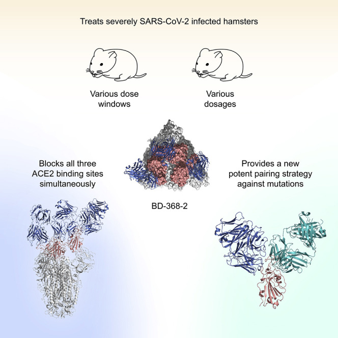
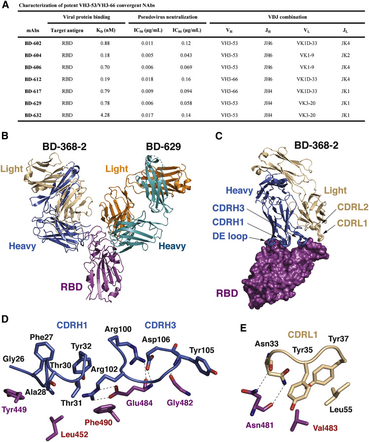
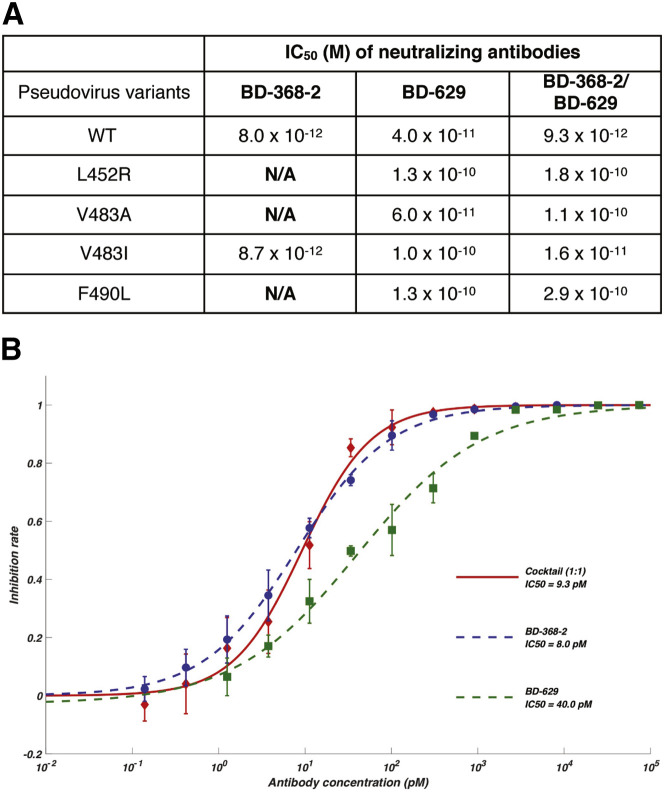
Understanding how potent neutralizing antibodies (NAbs) inhibit SARS-CoV-2 is critical for effective therapeutic development. We previously described BD-368-2, a SARS-CoV-2 NAb with high potency; however, its neutralization mechanism is largely unknown. Here, we report the 3.5-Å cryo-EM structure of BD-368-2/trimeric-spike complex, revealing that BD-368-2 fully blocks ACE2 recognition by occupying all three receptor-binding domains (RBDs) simultaneously, regardless of their "up" or "down" conformations. Also, BD-368-2 treats infected adult hamsters at low dosages and at various administering windows, in contrast to placebo hamsters that manifested severe interstitial pneumonia. Moreover, BD-368-2's epitope completely avoids the common binding site of VH3-53/VH3-66 recurrent NAbs, evidenced by tripartite co-crystal structures with RBDs. Pairing BD-368-2 with a potent recurrent NAb neutralizes SARS-CoV-2 pseudovirus at pM level and rescues mutation-induced neutralization escapes. Together, our results rationalized a new RBD epitope that leads to high neutralization potency and demonstrated BD-368-2's therapeutic potential in treating COVID-19.

Keywords: COVID-19; RBD; SARS-CoV-2; antibody cocktail; cryo-EM; epitope; hamster; mutation; neutralizing antibody; spike.


Conflict of interest statement

Declaration of Interests X.S.X. and Y.C. are inventors on the patent applications of the NAbs and co-founders of Singlomics Biopharmaceuticals. Other authors declare no competing financial interests.

Figures

None
Graphical abstract
Figure 1
Figure 1
Cryo-EM Structure of BD-368-2 Fabs in Complex with the S6P Trimer (A) BD-368-2 Fab induced drastic structural changes of S2P, as assessed by the negative stain EM, whereas S6P is more stable. The scale bar represents 100 nm. (B) Cryo-EM structure of the S6P trimer in complex with three BD-368-2 Fabs reconstructed at 3.5 Å. The three RBDs are highlighted in green (“up”) and yellow and magenta (“down”). The rest of the S trimer is shown in white. The fragment variable (Fv) region of BD-368-2 is shown in marine and wheat. (C) The structure of RBD in the RBD/BD-368-2 complex is overlaid onto the RBD in the RBD/ACE2/B0AT1 complex (PDB: 6M17). BD-368-2 would clash with both protomers in the ACE2 dimer and therefore interfere with the interaction between RBD and ACE2.
Figure S1
Figure S1
Workflow for the 3D Reconstruction of the Cryo-EM Structure of the S6P Trimer in Complex with Three BD-368-2 Fabs, Related to Figure 1 (A) Flow chart of image processing. (B) A representative raw image collected using a Titan Krios 300 kV equipped with a K2 detector. (C) Representative 2D classes. (D) Gold standard Fourier shell correlation (FSC) curve with the estimated resolution. (E) Eulerian angle distribution of the particles used in the final 3D reconstruction. (F) Local resolution estimation of the final density map analyzed by ResMap. (G) Representative density maps in the cryo-EM structure. Pro817, Pro892, and Pro899 (highlighted in red) are among the Pro substitutions in S6P that stabilizes the prefusion state structure.
Figure 2
Figure 2
Evaluation of BD-368-2’s In Vivo Efficacy at Various Dose Windows in the Adult Hamster Model (A) Experimental design for studying BD-368-2’s efficacy when administering at different time windows. Each group contains three hamsters. (B) Total weight loss, viral load, and pathological analyses for each hamster. Placebo hamsters were given PBS with equal volumes. The pathology score was determined based on the percentage of the lung section area that presented lesions. 1: 0–20%, 2: 20–40%, 3: 40–60%, 4: 60–80%, 5: 80–100%. Pneumonia severity was determined by H&E staining pathological analyses. 1: mild, 2: moderate, 3: severe. (C) The viral loads of the lung at day 7 determined by qRT-PCR (one-tailed t test, ∗∗p<0.01, n = 3, n represents the number of hamsters). Data are represented as mean ± SD. (D) Total body weight change (%) over 7 days. Data are represented as mean ± SD. (E) Weight change trajectory over 7 days.
Figure 3
Figure 3
Representative Pathological Evaluation of Lung Lesions for Study Groups with Different BD-368-2 Administering Windows (A) H&E staining of the lung section of C-1 in the placebo group. The histopathology image indicates severe interstitial pneumonia. (B) H&E staining of the lung section of P-3 in the −24 hpi group. The histopathology image indicates mild interstitial pneumonia. (C) H&E staining of the lung section of T1-2 in the 2 hpi group. The histopathology image indicates mild interstitial pneumonia. (D) H&E staining of the lung section of T2-3 in the 8 hpi group. The histopathology image indicates mild interstitial pneumonia. (E) H&E staining of the lung section of T3-1 in the 24 hpi group. The histopathology image indicates mild interstitial pneumonia. (F) H&E staining of the lung section of T4-2 in the 48 hpi group. The histopathology image indicates moderate-severe interstitial pneumonia.
Figure 4
Figure 4
Evaluation of BD-368-2’s In Vivo Efficacy at Various Dosages in the Adult Hamster Model (A) Experimental design for studying BD-368-2’s efficacy when administering different dosages at 2 hpi. Each group contains three hamsters. (B) Total weight loss, viral load, and pathological analyses for each hamster. Placebo hamsters were given PBS with equal volumes. Pathology score was determined based on the percentage of the lung section area that presented lesions. 1: 0–20%, 2: 20–40%, 3: 40–60%, 4: 60–80%, 5: 80–100%. Pneumonia severity was determined by H&E staining pathological analyses. 1: mild, 2: moderate, 3: severe. (C) The viral loads of the lung at day 7 determined by qRT-PCR (one-tailed t test, ∗∗p<0.01, p<0.05, n = 3, n represents the number of hamsters). Data are represented as mean ± SD. (D) Total body weight change (%) over 7 days. Data are represented as mean ± SD. (E) Weight change trajectory over 7 days.
Figure 5
Figure 5
Representative Pathological Evaluation of Lung Lesions for Study Groups with Different BD-368-2 Dosages (A) H&E staining of the lung section of C-2 in the placebo group. The histopathology image indicates severe interstitial pneumonia. (B) H&E staining of the lung section of T1-1 in the 20 mg/kg group. The histopathology image indicates mild interstitial pneumonia. (C) H&E staining of the lung section of T5-2 in the 10 mg/kg group. The histopathology image indicates mild interstitial pneumonia. (D) H&E staining of the lung section of T6-2 in the 5 mg/kg group. The histopathology image indicates moderate interstitial pneumonia. (E) H&E staining of the lung section of T7-2 in the 2 mg/kg group. The histopathology image indicates moderate-severe interstitial pneumonia.
Figure 6
Figure 6
Structures of the VH3-53/VH3-66 Antibodies in Tripartite Complexes with RBD and BD-368-2 (A) Characteristics of the potent VH3-53/VH3-66 recurrent NAbs selected based on VDJ sequences. (B) Crystal structure of RBD in complex with the Fabs of both BD-368-2 and BD-629. (C) Interaction between BD-368-2 Fab and RBD. BD-368-2 Fab is shown in ribbons, whereas RBD is shown in a surface view. The five regions in BD-368-2 that interact with RBD are highlighted using thicker ribbons. (D) Interactions between CDRH1, CDRH3, and RBD. Dashed lines indicate polar interactions. (E) Interactions between the DE loop in the BD-368-2 VH domain and RBD.
Figure S2
Figure S2
Binding Affinity and Neutralizing Abilities of VH3-53/VH3-66-Derived Antibodies, Related to Figure 6 (A) The distribution of IC50 against SARS-CoV-2 pseudovirus for VH3-53/VH3-66 derived antibodies revealed by high-throughput single-cell sequencing. Data for each antibody were obtained from a representative neutralization experiment, which contains three replicates. IC50 was calculated by using a four-parameter logistic curve-fitting and represented as mean. Each antibody’s heavy chain V-J gene is indicated on the x-axis, where the light chain V gene is indicated by different colors, as shown in the legend. A cross mark indicates that the antibody’s IC50 is higher than 1 μg/mL. The detailed characteristics of the antibodies shown here are listed in Table S2. (B) Neutralization potency measured by a SARS-CoV-2 spike-pseudotyped VSV neutralization assay. Data for each NAb were obtained from a representative neutralization experiment, which contains three replicates. Data are represented as mean ± SD. IC50 and IC80 were calculated by fitting a four-parameter logistic curve. (C) Measurement of the dissociation constant against RBD for the representing NAbs. All analyses were performed by using a serial 2-fold dilution of purified RBDs as the analyte, starting from 25 nM (magenta) to 1.56 nM (black).
Figure S3
Figure S3
The VH3-53/VH3-66 Antibodies Bind to RBD in a Similar Manner, Related to Figure 6 (A) Top: the crystal structure of BD-236 Fab in complex with RBD. The heavy chain (H) and light chain (L) of BD-236 Fab are shown in teal and orange, respectively. The RBD is shown in magenta. Disordered regions are depicted as dashed lines. Bottom: three heavy chain CDRs (CDRH1-3) and two light chain CDRs (CDRL1, CDRL3) in BD-236 Fab mediate the interaction with RBD. The CDRs are highlighted using thicker ribbons. RBD is shown in a surface view. (B) The crystal structure of BD-604 Fab in complex with RBD. (C) The crystal structure of BD-629 Fab in complex with RBD. (D) The crystal structures of other VH3-53/VH3-66 NAbs in complex with RBD. From left to right: B38 (PDB: 7BZ5), CB6 (PDB: 7C01), C105 (PDB: 6XCM), CV30 (PDB: 6XE1), CC12.1 (PDB: 6XC2), and CC12.3 (PDB: 6XC4).
Figure S4
Figure S4
Structures of the VH3-53/VH3-66 Antibodies in Tripartite Complexes with RBD and BD-368-2, Related to Figure 6 (A) Crystal structure of RBD in complex with the Fabs of both BD-368-2 and BD-236. (B) Crystal structure of RBD in complex with the Fabs of both BD-368-2 and BD-604. (C) Interaction between BD-629 and RBD in the BD-368-2/RBD/BD-629 tripartite complex. (D and E) Two aromatic clusters are critical for the interaction between BD-629 and RBD.
Figure 7
Figure 7
Potent BD-368-2/BD-629 Cocktail Rescues Mutation-Induced Neutralization Escapes (A) Individual NAbs and their combination were tested in mutated-pseudovirus neutralization assays. N/A indicates poor neutralization ability that IC50 could not be calculated. IC50 was calculated by fitting a four-parameter logistic curve. (B) Neutralization potency of BD-368-2/BD-629 cocktail on wild-type SARS-CoV-2 pseudotyped virus. Data were obtained from a representative neutralization experiment, which contains three replicates. Data are represented as mean ± SD.
Figure S5
Figure S5
Comparisons of the BD-368-2 and P2B-2F6, Related to Figure 6 (A) Structural comparison of BD-368-2 and P2B-2F6. RBD is shown in a surface view. The heavy chain and light chain of BD-368-2 Fab are shown in marine and wheat, respectively; whereas the heavy chain and light chain of P2B-2F6 Fab are shown in yellow and white. (B) P2B-2F6 would partially overlap with the VH3-53/VH3-66 antibodies such as BD-629, due to steric clashes between their VL domains. P2B-2F6 is shown in yellow and white, with its light chain highlighted using a surface view.
Figure S6
Figure S6
Comparisons of the BD-368-2/RBD/BD-629 Pair and REGN-COV2, Related to Figure 6 (A) Crystal structure of the BD-368-2/RBD/BD-629 complex. BD-368-2, RBD, and BD-629 are shown in marine, magenta, and teal, respectively. (B) Cryo-EM structure of the REGN10987/RBD/REGN10933 complex (PDB ID: 6XDG). REGN10987, RBD, and REGN10933 are shown in yellow, magenta, and light blue, respectively. (C) Structural superimposition of the above two complexes suggests that BD-629 can bind to RBD together with REGN10987, whereas BD-368-2 would clash with both REGN10987 and REGN10933 and therefore can’t function in a pair with any of them. (D) When a BD-368-2 molecule is bound to a “down” RBD in the S trimer, its constant domains may impose some steric hindrance for BD-629 to engage the “up” protomer. (E) A REGN10987 molecule interacting with a “down” RBD could also interfere with the binding of REGN10933 to the “up” RBD.
Figure S7
Figure S7
BD-368-2 Can Bind to the RBD Together with the VH3-53/VH3-66 Antibodies, S309, and CR3022, Related to Figure 6 The structures of the SARS-CoV-2 S in complex with S309 (PDB: 6WPS) and RBD in complex with CR3022 (PDB: 6W41) are superimposed onto the structure of BD-368-2/RBD/BD-629 to illustrate their binding modes. These antibodies have non-overlapping epitopes on RBD.

Similar articles

  • Potent neutralizing antibodies against multiple epitopes on SARS-CoV-2 spike.
    Liu L, Wang P, Nair MS, Yu J, Rapp M, Wang Q, Luo Y, Chan JF, Sahi V, Figueroa A, Guo XV, Cerutti G, Bimela J, Gorman J, Zhou T, Chen Z, Yuen KY, Kwong PD, Sodroski JG, Yin MT, Sheng Z, Huang Y, Shapiro L, Ho DD. Liu L, et al. Nature. 2020 Aug;584(7821):450-456. doi: 10.1038/s41586-020-2571-7. Epub 2020 Jul 22. Nature. 2020. PMID: 32698192
  • Mapping Neutralizing and Immunodominant Sites on the SARS-CoV-2 Spike Receptor-Binding Domain by Structure-Guided High-Resolution Serology.
    Piccoli L, Park YJ, Tortorici MA, Czudnochowski N, Walls AC, Beltramello M, Silacci-Fregni C, Pinto D, Rosen LE, Bowen JE, Acton OJ, Jaconi S, Guarino B, Minola A, Zatta F, Sprugasci N, Bassi J, Peter A, De Marco A, Nix JC, Mele F, Jovic S, Rodriguez BF, Gupta SV, Jin F, Piumatti G, Lo Presti G, Pellanda AF, Biggiogero M, Tarkowski M, Pizzuto MS, Cameroni E, Havenar-Daughton C, Smithey M, Hong D, Lepori V, Albanese E, Ceschi A, Bernasconi E, Elzi L, Ferrari P, Garzoni C, Riva A, Snell G, Sallusto F, Fink K, Virgin HW, Lanzavecchia A, Corti D, Veesler D. Piccoli L, et al. Cell. 2020 Nov 12;183(4):1024-1042.e21. doi: 10.1016/j.cell.2020.09.037. Epub 2020 Sep 16. Cell. 2020. PMID: 32991844 Free PMC article.
  • A Therapeutic Non-self-reactive SARS-CoV-2 Antibody Protects from Lung Pathology in a COVID-19 Hamster Model.
    Kreye J, Reincke SM, Kornau HC, Sánchez-Sendin E, Corman VM, Liu H, Yuan M, Wu NC, Zhu X, Lee CD, Trimpert J, Höltje M, Dietert K, Stöffler L, von Wardenburg N, van Hoof S, Homeyer MA, Hoffmann J, Abdelgawad A, Gruber AD, Bertzbach LD, Vladimirova D, Li LY, Barthel PC, Skriner K, Hocke AC, Hippenstiel S, Witzenrath M, Suttorp N, Kurth F, Franke C, Endres M, Schmitz D, Jeworowski LM, Richter A, Schmidt ML, Schwarz T, Müller MA, Drosten C, Wendisch D, Sander LE, Osterrieder N, Wilson IA, Prüss H. Kreye J, et al. Cell. 2020 Nov 12;183(4):1058-1069.e19. doi: 10.1016/j.cell.2020.09.049. Epub 2020 Sep 23. Cell. 2020. PMID: 33058755 Free PMC article.
  • Targeting SARS-CoV2 Spike Protein Receptor Binding Domain by Therapeutic Antibodies.
    Hussain A, Hasan A, Nejadi Babadaei MM, Bloukh SH, Chowdhury MEH, Sharifi M, Haghighat S, Falahati M. Hussain A, et al. Biomed Pharmacother. 2020 Oct;130:110559. doi: 10.1016/j.biopha.2020.110559. Epub 2020 Aug 1. Biomed Pharmacother. 2020. PMID: 32768882 Free PMC article. Review.
  • A Structural Landscape of Neutralizing Antibodies Against SARS-CoV-2 Receptor Binding Domain.
    Niu L, Wittrock KN, Clabaugh GC, Srivastava V, Cho MW. Niu L, et al. Front Immunol. 2021 Apr 28;12:647934. doi: 10.3389/fimmu.2021.647934. eCollection 2021. Front Immunol. 2021. PMID: 33995366 Free PMC article. Review.

Cited by

References

    1. Bao L., Deng W., Huang B., Gao H., Liu J., Ren L., Wei Q., Yu P., Xu Y., Qi F., et al. The pathogenicity of SARS-CoV-2 in hACE2 transgenic mice. Nature. 2020;583:830–833. - PubMed
    1. Barnes C.O., West A.P., Jr., Huey-Tubman K.E., Hoffmann M.A.G., Sharaf N.G., Hoffman P.R., Koranda N., Gristick H.B., Gaebler C., Muecksch F., et al. Structures of Human Antibodies Bound to SARS-CoV-2 Spike Reveal Common Epitopes and Recurrent Features of Antibodies. Cell. 2020;182:828–842.e16. - PMC - PubMed
    1. Baum A., Fulton B.O., Wloga E., Copin R., Pascal K.E., Russo V., Giordano S., Lanza K., Negron N., Ni M., et al. Antibody cocktail to SARS-CoV-2 spike protein prevents rapid mutational escape seen with individual antibodies. Science. 2020;369:1014–1018. - PMC - PubMed
    1. Brouwer P.J.M., Caniels T.G., van der Straten K., Snitselaar J.L., Aldon Y., Bangaru S., Torres J.L., Okba N.M.A., Claireaux M., Kerster G., et al. Potent neutralizing antibodies from COVID-19 patients define multiple targets of vulnerability. Science. 2020;369:643–650. - PMC - PubMed
    1. Callaway E., Cyranoski D., Mallapaty S., Stoye E., Tollefson J. The coronavirus pandemic in five powerful charts. Nature. 2020;579:482–483. - PubMed

Publication types

MeSH terms