. 2020 Oct:37:101725.
doi: 10.1016/j.redox.2020.101725. Epub 2020 Sep 15.

Glutathionylated and Fe-S cluster containing hMIA40 (CHCHD4) regulates ROS and mitochondrial complex III and IV activities of the electron transport chain

Affiliations

Glutathionylated and Fe-S cluster containing hMIA40 (CHCHD4) regulates ROS and mitochondrial complex III and IV activities of the electron transport chain

Venkata Ramana Thiriveedi et al. Redox Biol. 2020 Oct.

Abstract

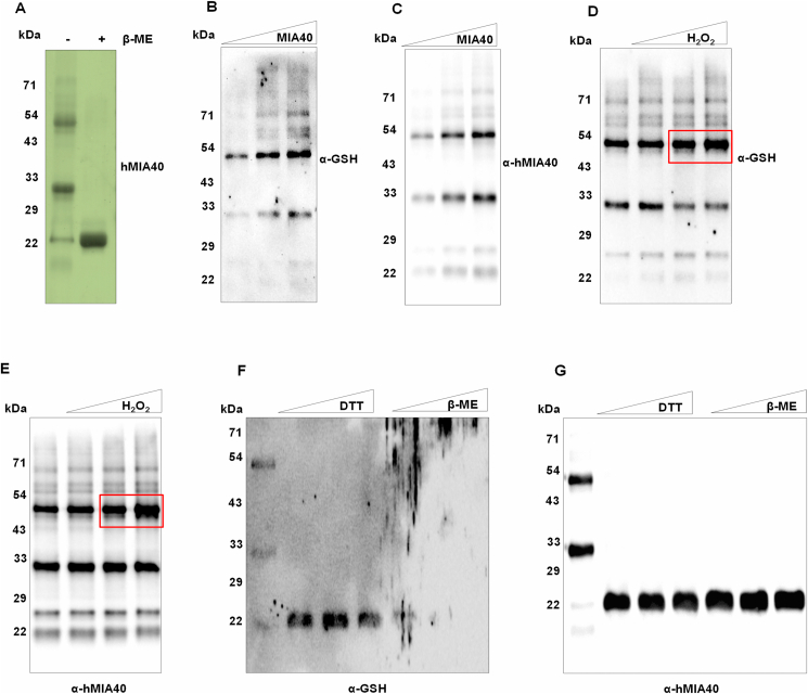
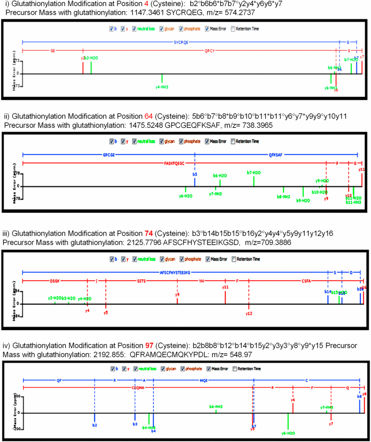
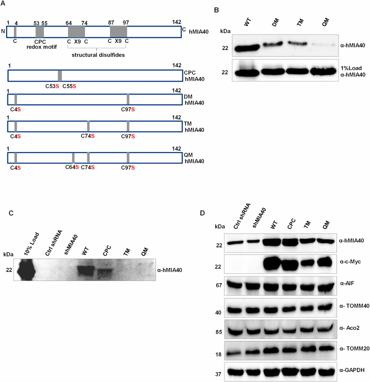
Human MIA40, an intermembrane space (IMS) import receptor of mitochondria harbors twin CX9C motifs for stability while its CPC motif is known to facilitate the import of IMS bound proteins. Site-directed mutagenesis complemented by MALDI on in vivo hMIA40 protein shows that a portion of MIA40 undergoes reversible S-glutathionylation at three cysteines in the twin CX9C motifs and the lone cysteine 4 residue. We find that HEK293T cells expressing hMIA40 mutant defective for glutathionylation are compromised in the activities of complexes III and IV of the Electron Transport Chain (ETC) and enhance Reactive Oxygen Species (ROS) levels. Immunocapture studies show MIA40 interacting with complex III. Interestingly, glutathionylated MIA40 can transfer electrons to cytochrome C directly. However, Fe-S clusters associated with the CPC motif are essential to facilitate the two-electron to one-electron transfer for reducing cytochrome C. These results suggest that hMIA40 undergoes glutathionylation to maintain ROS levels and for optimum function of complexes III and IV of ETC. Our studies shed light on a novel post-translational modification of hMIA40 and its ability to act as a redox switch to regulate the ETC and cellular redox homeostasis.

Keywords: Complex III and IV; Electron transport chain; Fe–S clusters; Glutathionylation; MIA40 (CHCHD4); Reactive oxygen species.


Conflict of interest statement

The authors declare that they have no conflicts of interest with the contents of this article.

Figures

Image 1
Graphical abstract
Fig. 1
Fig. 1
hMIA40 harbors reactive cysteines. (A) Recombinant and purified hMIA40 protein was treated with increasing concentration of DTT (1, 5, 10, and 20 mM) for 30 min before pull down with GSH-sepharose beads as described under Methods. After elution, GSH sepharose beads were resolved on SDS-PAGE and western blotted. Immunoblot probed with the hMIA40 antibody is shown. (B) It is the same as in (A) except that instead of DTT, increasing concentrations of H2O2 (1, 2, and 5 mM) used. (C) Lysate of HEK293T cells over-expressing hMIA40 were treated with increasing concentration of H2O2 (25, 50, and 100 μM) before pull-down with GSH-sepharose beads and processed as described in (A). Control indicates HEK293T cells transfected with vector. (D) Lysates (50 μg) from (C) before GSH pull-down were loaded on to SDS-PAGE and Western blotted. The immunoblot was probed with MIA40 and GAPDH antibodies to show the expression level of MIA40. (E) increasing concentrations (1 and 2 mg) of mitochondrial extracts from rat liver used for GSH pull-down assay and probed with MIA40 and ALR (F) Lysate of HEK293T cells over-expressing hMIA40 were treated with increasing concentration of NAC (1, 5 and 10 mM) for 12 h before immunoprecipitation with GSH antibody or pre-immune serum (PI) or no antibody (control) and binding to protein A sepharose beads. After resolution on SDS-PAGE and western blotting, the immunoblot probed with the hMIA40 antibody. (G) Lysates (50 μg) from (F) before immunoprecipitation resolved on SDS-PAGE, Western blotted, and the blots probed with antibodies against hMIA40, Aco1, Aco2, TOMM20, AIF, and GAPDH.
Fig. 2
Fig. 2
Glutathionylation of MIA40 is reversible. (A) Recombinant purified hMIA40 (10 μg) was resolved on a non-reducing and reducing SDS-PAGE and Coomassie-stained. (B&C) Increasing concentrations of recombinant hMIA40 (0.5, 1, & 1.5 μg) were resolved on a non-reducing SDS-PAGE, western transferred, and the blots probed with GSH and hMIA40 antibodies. (D&E) Same as in B&C, however, hMIA40 (10 μg) was treated with increasing concentrations of H2O2 (1, 2, and 5 mM) for 60 min before their resolution on a non-reducing SDS-PAGE. (F&G) Same as in D&E, however, hMIA40 (2 μg) was pre-treated with increasing concentrations of DTT (1, 5, and 10 mM) or β-mercaptoethanol (100, 150, and 200 mM) instead of H2O2.
Fig. 3
Fig. 3
Four cysteines in MIA40 are Glutathionylated. Lysate of HEK293T cells over-expressing Myc-His-hMIA40 was treated with 10 mM NAC before pull-down with GSH sepharose beads. After washing and elution, beads subjected to a non-reducing SDS-PAGE, and the gel Coomassie-stained. A 22 kDa band corresponding to hMIA40 kDa excised and subjected to MALDI analysis. LC-MS/MS analysis of gt cysteine peaks spectrum.
Fig. 4
Fig. 4
Validation of gt cysteines in hMIA40. (A) Schematic representation of the hMIA40 amino acid sequence shows the CPC and the dual CX9C motifs besides the seven cysteine positions. It additionally depicts the various cysteine to serine mutations generated by site-directed mutagenesis. (B) Recombinant hMIA40 wild type, DM, TM, and QM were purified from bacteria and used in GSH-sepharose pull-down as described in the Methods. Following SDS-PAGE and western blotting of the samples, the blots probed with the hMIA40 antibody. (C) HEK293T cells were co-transfected with scrambled shRNA and plasmid over-expressing Myc-His-hMIA40 wild type, CPC mutant, TM, or QM. Post-transfections, cells were treated with 10 mM NAC for 12 h before pull-down with GSH-sepharose beads. Samples resolved on SDS-PAGE, Western blotted, and the blots probed with hMIA40 antibody. (D) Lysates (50 μg) of transfected HEK293T cells from (C) were subjected to SDS-PAGE and western blotting before GSH-sepharose pull-down. The blots probed with antibodies against hMIA40, Myc, Aco2, TOMM20, TOMM40, AIF, and GAPDH.
Fig. 5
Fig. 5
Gt-hMIA40 regulates the ETC biogenesis and ROS production. Lysates of HEK293T transfected cells used as above. (A) Mitochondria were isolated, resolved on SDS-PAGE and western blotted. The blots probed with OXPHOS antibody that can detect significant components of ETC such as complex I (NDUFB8, 20 kDa), complex II (SDHB, 30 kDa), complex III (UQCRC2, 48 kDa), complex IV (MTCO1, 40 kDa) and complex V (ATP5A, 55 kDa) as shown (B) Quantification of blots from three independent experiments, (C, D, E & F) Activities of complexes I, II, III, and IV of ETC were analyzed spectrophotometrically as described in the Methods section and shown here. (G) ROS levels were measured in HEK293T transfected cells by H2DCFDA method, as described in the methods. All results plotted with mean ± S. E. (n = 3), *P ≤ 0.05. (H) Mitochondria of HEK293T cells over-expressing hMIA40 WT and QM mutant were used for immunocapture of complex III or pre-immune serum (PI) or no antibody (control) and binding to protein A sepharose beads. After resolution on SDS-PAGE and western blotting, the immunoblot probed with the Complex III antibody (H) and hMia40 and Cytochrome C (I).
Fig. 6
Fig. 6
hMIA40 reduces cytochrome C. (A) Oxidized cytochrome C (10 μM) was incubated with sodium dithionite in a buffer (50 mM potassium phosphate buffer pH7.4 and 0.5 mM EDTA) for 5 min before being scanned for absorption in the UV–visible spectral region. Shown here is a scan. (B) Recombinant hMIA40 wild type and mutant versions were over-expressed and affinity-purified from bacteria using Ni-NTA beads. 80 μg of the purified proteins were incubated with 10 μM oxidized cytochrome C as described in the Methods. The ability of hMIA40 wild type and mutant proteins to reduce oxidized cytochrome C was monitored in a Hitachi spectrophotometer by following the UV–visible spectra. (C) Oxidized cytochrome C incubated with 80 μg of wild type hMia40, CPC motif mutant hMIA40, and QM mutant hMIA40 in a buffer (50 mM potassium-phosphate buffer pH7.4) and cytochrome C reduction measured at 550 nm. (D) UV–Vis spectra of P1 (oligomer), P2 (dimer), and P3 (monomer) samples obtained after fractionation of his-hMia40. (E) Oxidized cytochrome C incubated with 80 μg of the oligomer, dimer, and monomer in a buffer (50 mM potassium-phosphate buffer pH7.4) and cytochrome C reduction measured at 550 nm. (F) Oxidized cytochrome C incubated with 80 μg of the oligomer, dimer and monomer in a buffer (50 mM potassium-phosphate buffer pH7.4 and 0.5 mM EDTA) and cytochrome C reduction measured at 550 nm. (G) UV–Vis spectra of wild type hMIA40, CPC mutant hMIA40, and QM mutant hMIA40 showing Fe–S absorption peak.
Fig. 7
Fig. 7
A proposed mechanistic scheme depicting the glutathionylation of MIA40 and transfer of electrons to cytochrome C. 1 & 2. ROS activates the reactive cysteines that subsequently get glutathionylated by GSH 3. ROS oxidizes GSH to GSSG. GSSG glutathionylates MIA40. 4. MIA40 gets deglutathionylated by the transfer of electrons to Fe–S motif present in CPC intramolecularly. 5. It accesses another CPC motif by dimerization/oligomerization in order to transfer another pair of electrons. The high molecular weight MIA40 gets reduced by transferring electrons to cytochrome C via Fe bound to CPC motif and monomerizes, and CPC motif forms disulfides. CPC motif reduces probably through protein import.

Similar articles

Cited by

References

    1. Deponte M., Hell K. Disulphide bond formation in the intermembrane space of mitochondria. J. Biochem. 2009;146:599–608. doi: 10.1093/jb/mvp133. - DOI - PubMed
    1. Hell K. The Erv1-Mia40 disulfide relay system in the intermembrane space of mitochondria. Biochim. Biophys. Acta. 2008;1783:601–609. doi: 10.1016/j.bbamcr.2007.12.005. - DOI - PubMed
    1. Mesecke N., Terziyska N., Kozany C., Baumann F., Neupert W., Hell K., Herrmann J.M. A disulfide relay system in the intermembrane space of mitochondria that mediates protein import. Cell. 2005;121:1059–1069. doi: 10.1016/j.cell.2005.04.011. - DOI - PubMed
    1. Finger Y., Riemer J. Protein import by the mitochondrial disulfide relay in higher eukaryotes. Biol. Chem. 2020;401:749–763. doi: 10.1515/hsz-2020-0108. - DOI - PubMed
    1. Habich M., Riemer J. Stop wasting protein—proteasome inhibition to target diseases linked to mitochondrial import. EMBO Mol. Med. 2019;11:e10441. doi: 10.15252/emmm.201910441. - DOI - PMC - PubMed

Publication types

MeSH terms

Substances

LinkOut - more resources