. 2020 Oct:60:102976.
doi: 10.1016/j.ebiom.2020.102976. Epub 2020 Sep 21.

Heterogeneous expression of the SARS-Coronavirus-2 receptor ACE2 in the human respiratory tract

Affiliations

Heterogeneous expression of the SARS-Coronavirus-2 receptor ACE2 in the human respiratory tract

Miguel E Ortiz et al. EBioMedicine. 2020 Oct.

Abstract

Background: Zoonotically transmitted coronaviruses are responsible for three disease outbreaks since 2002, including the current COVID-19 pandemic, caused by SARS-CoV-2. Its efficient transmission and range of disease severity raise questions regarding the contributions of virus-receptor interactions. ACE2 is a host ectopeptidase and the receptor for SARS-CoV-2. Numerous reports describe ACE2 mRNA abundance and tissue distribution; however, mRNA abundance is not always representative of protein levels. Currently, there is limited data evaluating ACE2 protein and its correlation with other SARS-CoV-2 susceptibility factors.

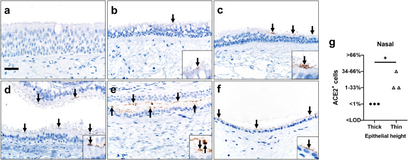
Materials and methods: We systematically examined the human upper and lower respiratory tract using single-cell RNA sequencing and immunohistochemistry to determine receptor expression and evaluated its association with risk factors for severe COVID-19.

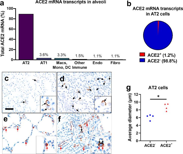
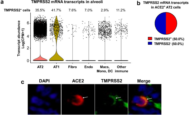
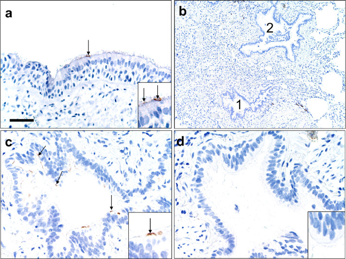
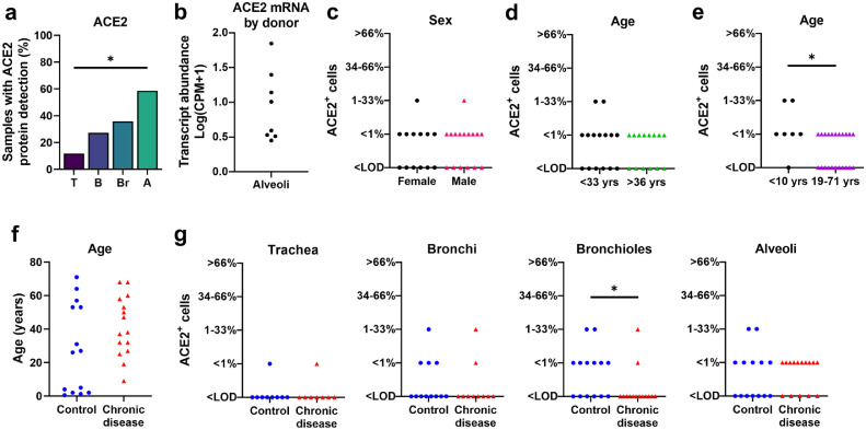
Findings: Our results reveal that ACE2 protein is highest within regions of the sinonasal cavity and pulmonary alveoli, sites of presumptive viral transmission and severe disease development, respectively. In the lung parenchyma, ACE2 protein was found on the apical surface of a small subset of alveolar type II cells and colocalized with TMPRSS2, a cofactor for SARS-CoV2 entry. ACE2 protein was not increased by pulmonary risk factors for severe COVID-19. Additionally, ACE2 protein was not reduced in children, a demographic with a lower incidence of severe COVID-19.

Interpretation: These results offer new insights into ACE2 protein localization in the human respiratory tract and its relationship with susceptibility factors to COVID-19.

Keywords: Alveolar type II cells; Ciliated cells; Expression; Immunohistochemistry; Lung.


Conflict of interest statement

Declaration of Competing Interest The authors declare no competing interests related to this work. This work was supported by the National Institutes of Health (NIH, P01 AI060699). P.B.M. is on the scientific advisory board and receives support for sponsored research from Spirovant Sciences, Inc. P.B.M. is on the scientific advisory board for Oryn Therapeutics.

Figures

Fig 1
Fig. 1
ACE2 expression in human lung. a, b) Single-cell RNA sequencing reanalysis of ACE2 transcript abundance in alveoli from lung parenchyma samples . Summative observations from all donors. Airway cells (basal, mitotic, ciliated, club) are not shown. a) 89•5% of the cells with detectable ACE2 mRNA in the alveoli are alveolar type II cells. b) Only 1•2% of alveolar type II cells have ACE2 mRNA transcripts. c-f) Detection of ACE2 protein (brown color, black arrows, and insets) in representative sections of lower respiratory tract regions and tissue scoring (see Supplemental Table 2) (g). c, d) Alveolar regions had uncommon to regional polarized apical staining of solitary epithelial cells (c) that (when present) were more readily detected in collapsed regions of lung (d). e, f) SP-C (red arrows, inset) and ACE2 (black arrows, inset) dual immunohistochemistry on the same tissue sections. e) Non-collapsed regions had normal SP-C+ AT2 cells lacking ACE2. f) Focal section of peri-airway remodeling and collapse with several SP-C+ (red arrows) AT2 cells, but only a small subset of AT2 cells had prominent apical ACE2 protein (black arrows, inset). g) SP-C+/ACE2+ AT2 cells were often larger than SP-C+/ACE2 AT2 cells from same lung sections (see also d and e insets) indicative of AT2 hypertrophy, each data point represents the average value for each case from 5-10 cell measurements per group, P=0•0014, paired T-test. AT2: alveolar type II. AT1: alveolar type I. Macs: Macrophages. Mono: Monocytes. DC: dendritic cells. Other immune cells: B cells, mast cells, natural killer/T cells. Endo: Endothelial. Fibro: Fibroblasts/myofibroblasts. Bar = 35 μm.
Fig 2
Fig. 2
TMPRSS2 expression in the alveoli. a, b) Single-cell RNA sequencing reanalyses of TMPRSS2 transcript abundance in alveoli from lung parenchyma . Summative observations from all donors. a) Percentage of TMPRSS2+ cells within each cell type shows TMPRSS2 transcripts in 35•5% of alveolar type II cells. Airway cells (basal, mitotic, ciliated, club) are not shown. Violin plots represent expression, each data point denotes a cell. b) TMPRSS2 transcripts in ACE2+ alveolar type II cells. c) Immunofluorescence of alveoli shows apical colocalization of ACE2 and TMPRSS2 (white arrows). AT2: alveolar type II. AT1: alveolar type I. Macs: Macrophages. Mono: Monocytes. DC: dendritic cells. Other immune cells: B cells, mast cells, natural killer/T cells. Endo: Endothelial. Fibro: Fibroblasts/myofibroblasts. CPM: counts per million.
Fig 3
Fig. 3
ACE2 protein in human lower airways. a) Large airways (trachea and bronchi) exhibited rare ACE2 protein on the apical surface of ciliated cells. b-d) Small airways (bronchioles) exhibited uncommon to localized apical ACE2 protein in ciliated cells (c, #1 in b) while the adjacent bronchioles (d, #2 in b) lacked protein. Bar = 35 (a), 140 (b), and 70 μm (c, d).
Fig 4
Fig. 4
ACE2 distribution and scores in respiratory tissues. a) ACE2 protein had progressively increased detection between donors in tissues from trachea (T), bronchi (B), bronchioles (Br), to alveoli (A), (P=0•0009, Cochran-Armitage test for trend). b) Single-cell RNA sequencing reanalyses of ACE2 mRNA transcript abundance in the alveoli shows variation between donors . c-d) ACE2 protein scores from lung donors showed no differences based on sex or lower vs. upper ages (using median age as a cut-off) (A, P=0•7338 and B, P=0•7053, Mann-Whitney U test). e) ACE2 protein scores were elevated in young children (<10 yrs) compared to the remaining subjects (19-71 yrs) (P=0•0282 Mann-Whitney U test). f) Control and chronic disease groups did not have any significant differences in age (P=0•1362 Mann-Whitney U test). g) ACE2 protein scores for trachea, bronchi, bronchiole, and alveoli in control versus chronic disease groups (P= >0•9999, 0•6263, 0•0433, and 0•7359, respectively, Mann-Whitney U test). Each symbol represents one donor. CPM: counts per million. LOD: Limit of detection.
Fig 5
Fig. 5
ACE2 protein in sinonasal tissues. Detection of ACE2 protein (brown color, arrows, and insets, a-f) and tissue scoring (g) in representative sections of nasal tissues. a, b) In thick pseudostratified epithelium (PSE) ACE2 protein was absent (a) to rare (b) and apically located on ciliated cells. c) Tissue section shows a transition zone from thick (left side, > ~4 nuclei) to thin (right side, ≤ ~4 nuclei) PSE and ACE2 protein was restricted to the apical surface of the thin PSE. d-f) ACE2 protein was detected multifocally on the apical surface of ciliated cells in varying types of thin PSE, even to simple cuboidal epithelium (f). Bar = 30 μm. g) ACE2 protein detection scores for each subject were higher in thin than thick epithelium, (P=0•05, Mann-Whitney U test). LOD: Limit of detection.

Update of

Comment in

References

    1. Li W, Moore MJ, Vasilieva N, Sui J, Wong SK, Berne MA. Angiotensin-converting enzyme 2 is a functional receptor for the SARS coronavirus. Nature. 2003;426(6965):450–454. - PMC - PubMed
    1. Zhou P, Yang XL, Wang XG, Hu B, Zhang L, Zhang W. A pneumonia outbreak associated with a new coronavirus of probable bat origin. Nature. 2020;579(7798):270–273. - PMC - PubMed
    1. Lam CW, Chan MH, Wong CK. Severe acute respiratory syndrome: clinical and laboratory manifestations. Clin Biochem Rev. 2004;25(2):121–132. - PMC - PubMed
    1. Wilder-Smith A, Chiew CJ, Lee VJ. Can we contain the COVID-19 outbreak with the same measures as for SARS? Lancet Infect Dis. 2020;20(5):e102–e107. - PMC - PubMed
    1. Zhou F, Yu T, Du R, Fan G, Liu Y, Liu Z. Clinical course and risk factors for mortality of adult inpatients with COVID-19 in Wuhan, China: a retrospective cohort study. Lancet. 2020;395(10229):1054–1062. - PMC - PubMed

MeSH terms