

Use of 'U-shaped tool for follow up of corneal ulcer cases in the COVID-19 pandemic
- PMID: 32971640
- PMCID: PMC7728037
- DOI: 10.4103/ijo.IJO_1560_20
Use of 'U-shaped tool for follow up of corneal ulcer cases in the COVID-19 pandemic
Abstract
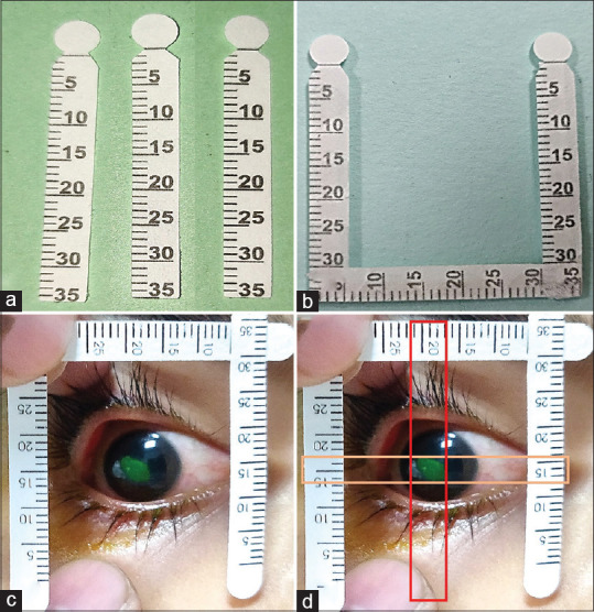
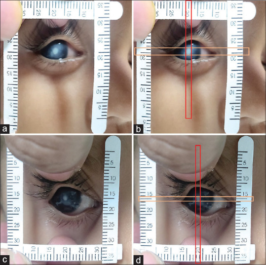
To describe a smartphone-based telemedicine tool for monitoring of corneal ulcer size during the corona pandemic, a simple "U"-shaped tool was constructed using three Schirmer's strips that were provided to the patients with small to medium-sized corneal ulcers. The patient and the attendant were trained to use this simple U-shaped tool at home and send digital images to the treating ophthalmologist, to monitor the course of the ulcer. The tool was used in five eyes of five patients with active microbial keratitis. Patients were followed up regularly with the use of telemedicine facility every 48 h for an average duration of 7.6 days (range 6-9 days). In all the five eyes, assessment of the serial images with U-shaped tool showed decrease in size of corneal ulcer, which corroborated with subjective improvement in symptoms. Hence, the novel "'U'-shaped tool" may provide an effective measure in following-up of corneal ulcer patients in times of the COVID-19 pandemic, obviating frequent hospital visits and risk of contracting COVID.
Keywords: Bacterial keratitis; COVID-19; corneal ulcer; follow-up; pandemic.
Conflict of interest statement
None
Figures
Similar articles
-
Smartphone assisted slit lamp evaluation during the COVID-19 pandemic.Indian J Ophthalmol. 2020 Jul;68(7):1492. doi: 10.4103/ijo.IJO_1653_20. Indian J Ophthalmol. 2020. PMID: 32587214 Free PMC article. No abstract available.
-
Access to Telemedicine-Are We Doing All That We Can during the COVID-19 Pandemic?Otolaryngol Head Neck Surg. 2020 Jul;163(1):104-106. doi: 10.1177/0194599820925049. Epub 2020 May 5. Otolaryngol Head Neck Surg. 2020. PMID: 32366162 Review.
-
Monitoring and Management of Home-Quarantined Patients With COVID-19 Using a WeChat-Based Telemedicine System: Retrospective Cohort Study.J Med Internet Res. 2020 Jul 2;22(7):e19514. doi: 10.2196/19514. J Med Internet Res. 2020. PMID: 32568727 Free PMC article.
-
Health Care After the COVID-19 Pandemic and the Influence of Telemedicine.Mayo Clin Proc. 2020 Sep;95(9S):S66-S68. doi: 10.1016/j.mayocp.2020.06.052. Epub 2020 Jul 27. Mayo Clin Proc. 2020. PMID: 32948262 Free PMC article. Review. No abstract available.
-
Telemedicine in neurosurgery during the novel coronavirus (COVID-19) pandemic.Neurol Neurochir Pol. 2020;54(2):207-208. doi: 10.5603/PJNNS.a2020.0038. Epub 2020 Apr 22. Neurol Neurochir Pol. 2020. PMID: 32319670 No abstract available.
Cited by
-
Publication trend of COVID-19 and non-COVID-19 articles in the Indian Journal of Ophthalmology during the pandemic.Indian J Ophthalmol. 2021 May;69(5):1241-1248. doi: 10.4103/ijo.IJO_117_21. Indian J Ophthalmol. 2021. PMID: 33913868 Free PMC article.
-
A Situational Analysis of the Impact of the COVID-19 Pandemic on Digital Health Research Initiatives in South Asia.Cureus. 2023 Nov 17;15(11):e48977. doi: 10.7759/cureus.48977. eCollection 2023 Nov. Cureus. 2023. PMID: 38111408 Free PMC article. Review.
-
Validating the use of U-tool as a novel method for measuring the corneal diameter in infants screened for congenital glaucoma.Indian J Ophthalmol. 2022 Jan;70(1):143-146. doi: 10.4103/ijo.IJO_930_21. Indian J Ophthalmol. 2022. PMID: 34937226 Free PMC article.
References
-
- Kim RY, Cooper KL, Kelly LD. Predictive factors for response to medical therapy in bacterial ulcerative keratitis. Graefes Arch Clin Exp Ophthalmol. 1996;234:731–8. - PubMed
-
- Mukerji N, Vajpayee RB, Sharma N. Technique of area measurement of epithelial defects. Cornea. 2003;22:549–51. - PubMed
-
- Mazumder H, Hossain MM, Das A. Geriatric care during public health emergencies: Lessons learned from novel corona virus disease (COVID-19) pandemic. J Gerontol Soc Work. 2020;63:257–8. - PubMed
MeSH terms
LinkOut - more resources
Full Text Sources
Medical
