

Smartphone-based intraocular lens microscope
- PMID: 32971646
- PMCID: PMC7727942
- DOI: 10.4103/ijo.IJO_2032_19
Smartphone-based intraocular lens microscope
Abstract
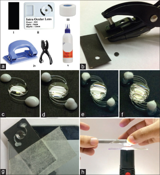
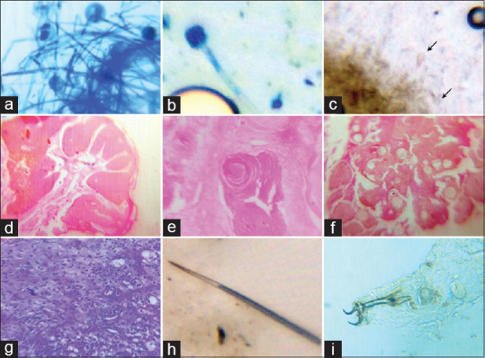
Microscopes play an important role in the diagnosis of microorganisms and pathological lesions in ophthalmology guiding us to the appropriate management. The current trend of collecting samples and examination is mostly laboratory-based which consume time, labor, and are costly. Smartphones are being used in different fields of ophthalmology with great ubiquity. The good quality photographs obtained by smartphones along with the ease of mobility has made it possible to warrant its use in the microscopic world. This article describes a simple novel technique of preparing an intraocular lens system which can be used in conjunction with a smartphone to detect microorganisms and pathological lesions.
Keywords: Innovation; low-cost device; microscope; point of care diagnosis; smartphone photography.
Conflict of interest statement
None
Figures
Similar articles
-
Smartphone Gonioscopy With a Magnifying Intraocular Lens: A Cost-effective Angle Imaging Device.J Glaucoma. 2022 May 1;31(5):356-360. doi: 10.1097/IJG.0000000000002006. Epub 2022 Feb 28. J Glaucoma. 2022. PMID: 35220386
-
Anterior segment photography with intraocular lens.Indian J Ophthalmol. 2019 Oct;67(10):1690-1691. doi: 10.4103/ijo.IJO_52_19. Indian J Ophthalmol. 2019. PMID: 31546510 Free PMC article. No abstract available.
-
Slit-lamp based intraocular lens microscope - A novel technique of rapid office-based microscopy.Indian J Ophthalmol. 2022 Apr;70(4):1381-1383. doi: 10.4103/ijo.IJO_2389_21. Indian J Ophthalmol. 2022. PMID: 35326059 Free PMC article.
-
Clinically useful smartphone ophthalmic imaging techniques.Graefes Arch Clin Exp Ophthalmol. 2021 Feb;259(2):279-287. doi: 10.1007/s00417-020-04917-z. Epub 2020 Sep 11. Graefes Arch Clin Exp Ophthalmol. 2021. PMID: 32915278 Review.
-
Intraocular lens centration and stability: efficacy of current technique and technology.Curr Opin Ophthalmol. 2009 Jan;20(1):33-6. doi: 10.1097/icu.0b013e328318591c. Curr Opin Ophthalmol. 2009. PMID: 19093329 Review.
Cited by
-
Innovative rapid detection of ocular surface parasitosis in under-resourced facilities.Indian J Ophthalmol. 2022 Aug;70(8):3153-3154. doi: 10.4103/ijo.IJO_666_22. Indian J Ophthalmol. 2022. PMID: 35918999 Free PMC article. No abstract available.
-
A corneal stroma circular ring captured by smartphone adaptor slit lamp camera after small incision lenticule extraction.Clin Case Rep. 2024 Mar 31;12(4):e8690. doi: 10.1002/ccr3.8690. eCollection 2024 Apr. Clin Case Rep. 2024. PMID: 38562576 Free PMC article.
-
Commentary: What the eye sees, Let's make the world see - Smart evolution of teleophthalmology.Indian J Ophthalmol. 2022 Dec;70(12):4243-4244. doi: 10.4103/ijo.IJO_2116_22. Indian J Ophthalmol. 2022. PMID: 36453324 Free PMC article. No abstract available.
-
Fungal corneal ulcer detection in a slit-lamp-based intraocular lens microscopy clinical setup.Indian J Ophthalmol. 2023 May;71(5):2301-2302. doi: 10.4103/IJO.IJO_65_23. Indian J Ophthalmol. 2023. PMID: 37202985 Free PMC article. No abstract available.
-
Trends in Ophthalmological Patents, 2005-2020.J Ocul Pharmacol Ther. 2023 Jul;39(6):365-370. doi: 10.1089/jop.2022.0185. Epub 2023 May 16. J Ocul Pharmacol Ther. 2023. PMID: 37192496 Free PMC article.
References
-
- Anselmi F, Grier Z, Soddu M, Kenyatta N, Odame S, Sanders J, et al. A low-cost do-it-yourself microscope kit for hands-on science education. Optics Education and Outreach V. 2018
-
- Keller E, Goldman R. Light Microscopy. Woodbury, NY: Cold Spring Harbor Laboratory Press; 2006. p. 8.
-
- Bozzola JJ, Russell LD. Electron Microscopy: Principles and Techniques for Biologists. Sudbury MA: Jones and Bartlett Learning; 1999. pp. 2–16.
-
- Cheng PC, Jan GJ. X-ray Microscopy: Instrumentation and Biological Applications. Berlin Heidelberg: Springer Science and Business Media; 2012. pp. 1–12.
-
- Muller M. Introduction to Confocal Fluorescence Microscopy. Bellingham WA: SPIE Press; 2006. pp. 1–23.
MeSH terms
LinkOut - more resources
Full Text Sources
