Review
. 2020 Sep 22;9(9):2138.
doi: 10.3390/cells9092138.

Oxidoreductases in Glycoprotein Glycosylation, Folding, and ERAD

Affiliations
Review

Oxidoreductases in Glycoprotein Glycosylation, Folding, and ERAD

Chaitanya Patel et al. Cells. .

Abstract

N-linked glycosylation and sugar chain processing, as well as disulfide bond formation, are among the most common post-translational protein modifications taking place in the endoplasmic reticulum (ER). They are essential modifications that are required for membrane and secretory proteins to achieve their correct folding and native structure. Several oxidoreductases responsible for disulfide bond formation, isomerization, and reduction have been shown to form stable, functional complexes with enzymes and chaperones that are involved in the initial addition of an N-glycan and in folding and quality control of the glycoproteins. Some of these oxidoreductases are selenoproteins. Recent studies also implicate glycan machinery-oxidoreductase complexes in the recognition and processing of misfolded glycoproteins and their reduction and targeting to ER-associated degradation. This review focuses on the intriguing cooperation between the glycoprotein-specific cell machineries and ER oxidoreductases, and highlights open questions regarding the functions of many members of this large family.

Keywords: ER quality control; ERAD; PDI; calnexin; mannosidase; oligosaccharyltransferase.


Conflict of interest statement

The authors declare no conflict of interest.

Figures

Figure 1
Figure 1
Glycoprotein translocation and folding in the endoplasmic reticulum (ER) in mammalian cells. (1) The oligosaccharide transferase (OST) complex co-translationally transfers the N-glycan precursor (Glc3Man9GlcNAc2) from the lipid-linked oligosaccharide (LLO) (Glc3Man9GlcNAc2-PP-Dolichol) to an asparagine residue in an N-glycosylation sequon of the glycoprotein during its translocation into the ER. (2) Sugar chain transfer to the glycoprotein is assisted by mixed disulfide bond formation between the glycoprotein and MAGT1 (IAP) or TUSC3 (N33) (in complex with the catalytic OST subunit STT3B), or with selenoprotein T (SELENOT) (in complex with the catalytic OST subunit STT3A). The names of other subunits of the large OST complex are omitted for simplicity. (3) The two terminal glucose residues are removed by Glucosidase I and II (GI and GII), respectively, allowing the glycoprotein to enter folding cycles by the ER chaperones calnexin (CNX) and calreticulin (CRT). (4) The oxidoreductase ERp57 interacts with CNX and CRT, assisting disulfide bond formation. Similar activity is observed by ERp72 indirectly interacting with CRT via PDIR. (5) The removal of the last glucose residue by GII results in the exit of the glycoprotein from the CNX cycle. (6) Properly folded glycoproteins are sent to the Golgi via the ER–Golgi intermediate compartment (ERGIC), mediated by a complex of ERGIC-53 and the oxidoreductase ERp44. (7) The ERGIC53/ERp44 complex is shuttled via COPII vesicles to the ERGIC, where it aids glycoprotein oligomerization. Unassembled subunits are shuttled back to the ER with ERp44 via COPI vesicles. (8) Misfolded glycoproteins are retained in the ER, where they are recognized by UDPGlc:glycoprotein glucosyltransferase (UGGT), with its 4 inactive Trxl domains in a complex with the seleno-oxidoreductase Sep15. UGGT reglucosylates the glycoprotein for re-entry into the CNX folding cycle. (9) Terminally misfolded glycoproteins are targeted to ER-associated degradation (ERAD). Oxidoreductases are marked in red.
Figure 2
Figure 2
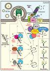
N-Glycan processing and ERAD in mammalian cells. (1) Misfolded glycoproteins carrying Man9GlcNAc2 are targeted to the ER-derived quality control compartment (ERQC), and their N-glycans are subjected to mannose trimming for their recognition by the ERAD machinery. A scheme on the right depicts mannose trimming of α1,2 mannose residues (light green circles, other mannoses are in dark green, and blue squares are GlcNAc). (2–5) This processing is performed sequentially or in parallel by the α1,2 mannosidases EDEM1-3, ManIA, or ERmanI. (2) ManIA or ERmanI (and possibly the EDEMs) reside in quality control vesicles (QCVs) and interact with the misfolded glycoproteins, likely at vesicle fusion sites. Several oxidoreductases interact with the EDEMs–(3) PDI (PDA1, P4HB) and (5) TXNDC11 associate with EDEM1 and 2, while TXNDC11 and (4) ERp46 (TXNDC5) associate with EDEM3. The associations of the oxidoreductases accelerate the mannose trimming activity of the EDEMs on misfolded glycoproteins, as shown so far for PDI and TXNDC11 with EDEM1/2 and ERp46 with EDEM3. (6,7) Trimming of three or all four α1,2 mannose residues results in glycoprotein molecules carrying Man5-6GlcNAc2 N-glycans, which bind to the OS-9/XTP3B lectins that interact with the ERAD complex. (7) OS-9 interacts with SEL1L, an adaptor protein for the E3 ubiquitin ligase HRD1, which associates with the membrane proteins Herp and the Derlins 1–3, among others. (8) OS-9 and SEL1L also interact with a complex of proteins containing Trxl domains, the non-catalytic ERp90 (TXNDC16), and the reductase ERdj5. ERp90 associates with the NAPDH-dependent reductase ERFAD. ERdj5 also interacts with EDEM1. These complexes lead to misfolded glycoprotein reduction, polyubiquitination, and retrotranslocation, with the help of the AAA ATPase p97 on the cytosolic side. (9) Peptide:N-glycosidase (PNGase) removes the N-glycans prior to deubiquitylation and degradation of the misfolded glycoproteins by the proteasomes. For simplicity, several ERAD components, such as p97 adaptors and proteasome shuttling factors, are not depicted.

References

    1. Bošnjak I., Bojović V., Šegvić-Bubić T., Bielen A. Occurrence of protein disulfide bonds in different domains of life: A comparison of proteins from the Protein Data Bank. Protein. Eng. Des. Sel. 2014;27:65–72. doi: 10.1093/protein/gzt063. - DOI - PubMed
    1. Robinson P.J., Bulleid N.J. Mechanisms of Disulfide Bond Formation in Nascent Polypeptides Entering the Secretory Pathway. Cells. 2020;9:1994. doi: 10.3390/cells9091994. - DOI - PMC - PubMed
    1. Guerra C., Molinari M. Thioredoxin-Related Transmembrane Proteins: TMX1 and Little Brothers TMX2, TMX3, TMX4 and TMX5. Cells. 2020;9:2000. doi: 10.3390/cells9092000. - DOI - PMC - PubMed
    1. Araki K., Inaba K. Structure, mechanism, and evolution of Ero1 family enzymes. Antioxid. Redox. Signal. 2012;16:790–799. doi: 10.1089/ars.2011.4418. - DOI - PubMed
    1. Gross E., Sevier C.S., Heldman N., Vitu E., Bentzur M., Kaiser C.A., Thorpe C., Fass D. Generating disulfides enzymatically: Reaction products and electron acceptors of the endoplasmic reticulum thiol oxidase Ero1p. Proc. Natl. Acad. Sci. USA. 2006;103:299–304. doi: 10.1073/pnas.0506448103. - DOI - PMC - PubMed

Publication types

MeSH terms

LinkOut - more resources