. 2020 Sep 22;8(9):369.
doi: 10.3390/biomedicines8090369.

Chronic Electrical Stimulation of the Superior Laryngeal Nerve in the Rat: A Potential Therapeutic Approach for Postmenopausal Osteoporosis

Affiliations

Chronic Electrical Stimulation of the Superior Laryngeal Nerve in the Rat: A Potential Therapeutic Approach for Postmenopausal Osteoporosis

Kaori Iimura et al. Biomedicines. .

Abstract

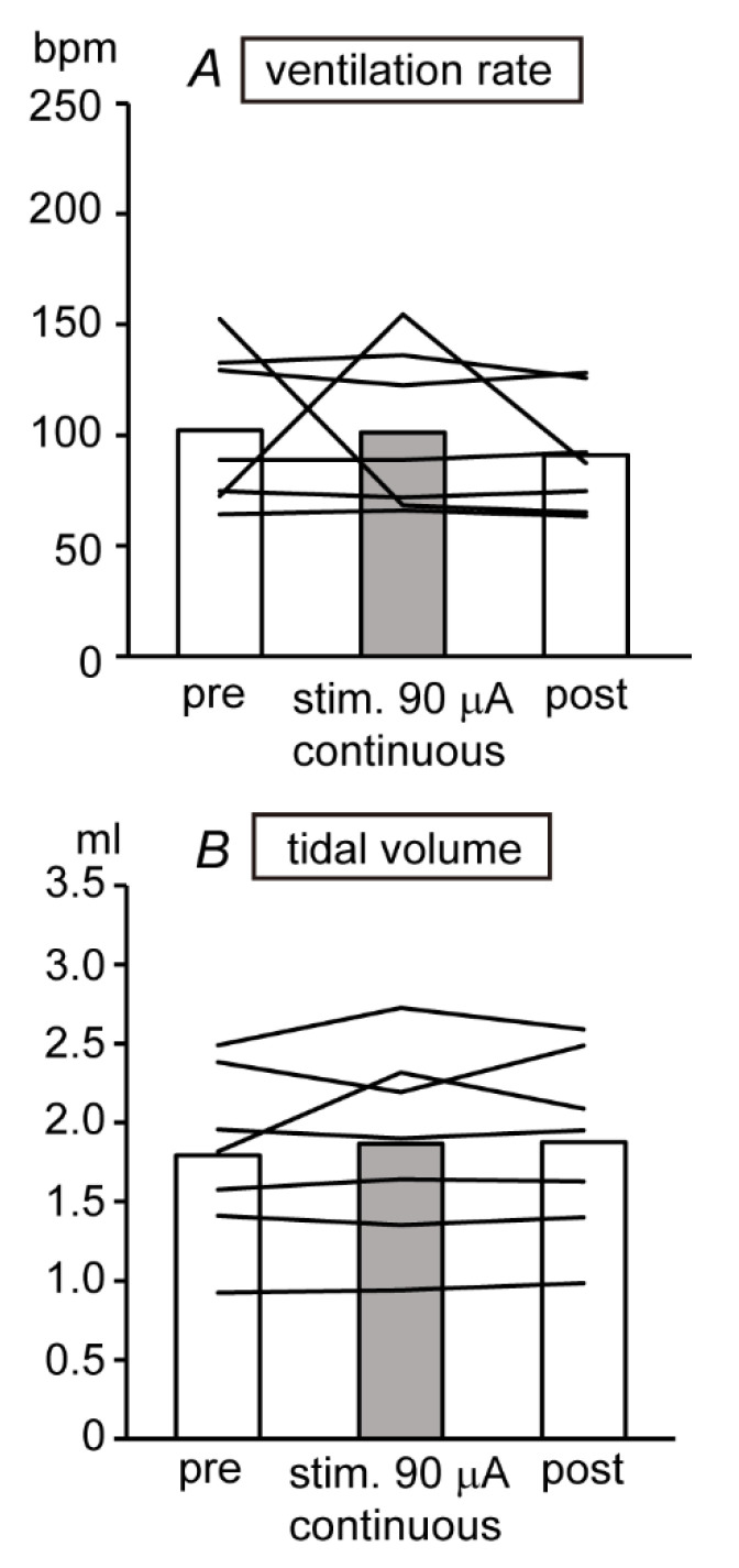
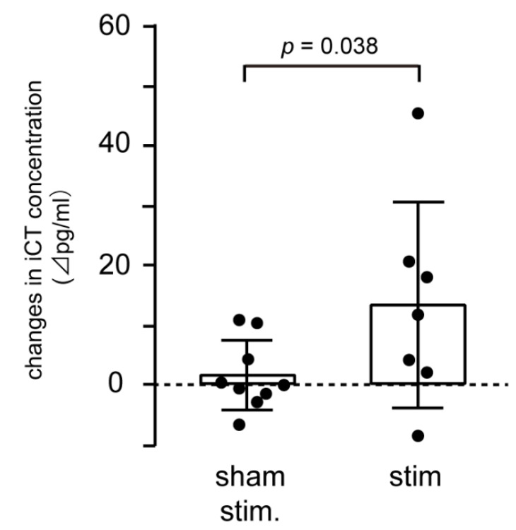
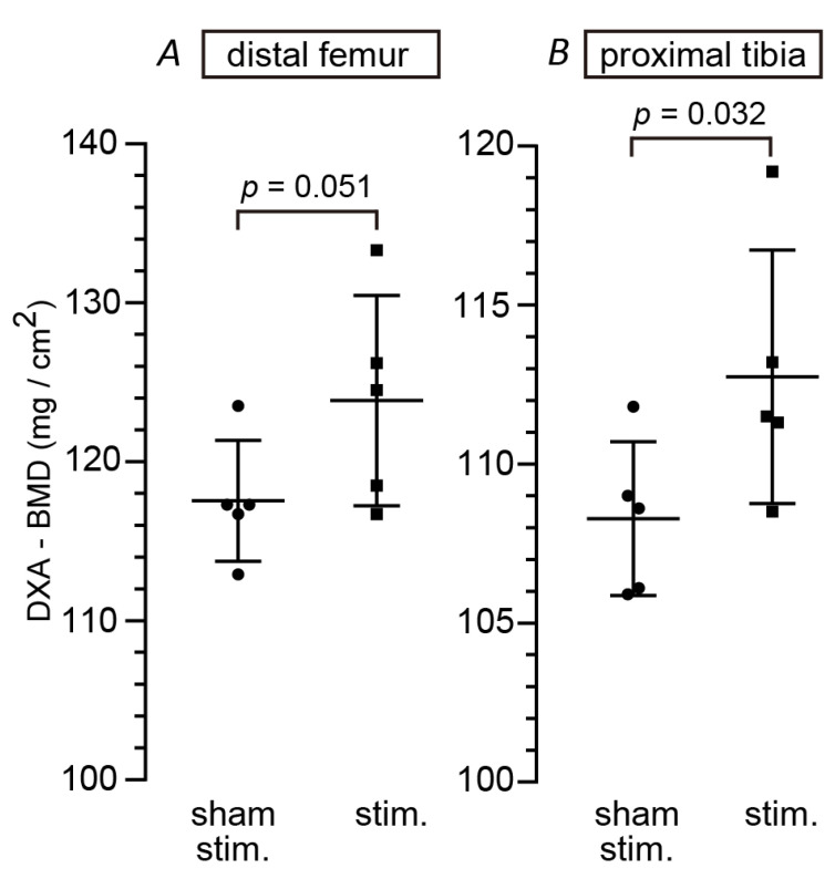
Electrical stimulation of myelinated afferent fibers of the superior laryngeal nerve (SLN) facilitates calcitonin secretion from the thyroid gland in anesthetized rats. In this study, we aimed to quantify the electrical SLN stimulation-induced systemic calcitonin release in conscious rats and to then clarify effects of chronic SLN stimulation on bone mineral density (BMD) in a rat ovariectomized disease model of osteoporosis. Cuff electrodes were implanted bilaterally on SLNs and after two weeks recovery were stimulated (0.5 ms, 90 microampere) repetitively at 40 Hz for 8 min. Immunoreactive calcitonin release was initially measured and quantified in systemic venous blood plasma samples from conscious healthy rats. For chronic SLN stimulation, stimuli were applied intermittently for 3-4 weeks, starting at five weeks after ovariectomy (OVX). After the end of the stimulation period, BMD of the femur and tibia was measured. SLN stimulation increased plasma immunoreactive calcitonin concentration by 13.3 ± 17.3 pg/mL (mean ± SD). BMD in proximal metaphysis of tibia (p = 0.0324) and in distal metaphysis of femur (p = 0.0510) in chronically SLN-stimulated rats was 4-5% higher than that in sham rats. Our findings demonstrate chronic electrical stimulation of the SLNs produced enhanced calcitonin release from the thyroid gland and partially improved bone loss in OVX rats.

Keywords: bone mineral density; calcitonin; electrical stimulation; neuromodulation; osteoporosis; ovariectomy; superior laryngeal nerve; thyroid gland.


Conflict of interest statement

The funder was involved in study design, project management, the decision to publish the results, and final approval of manuscript.

Figures

Figure 1
Figure 1
Time schedule of Experiment 2. Rats received ovariectomy (OVX) surgery at 12 weeks of age. They received another surgery of implanting cuff electrodes on the superior laryngeal nerves (SLNs) and fixing a head socket on the skull at three weeks after OVX. SLNs stimulation was started at five weeks after OVX and continued for a period of 3–4 weeks (8 min/h × 7 times/day × 4 days/week). After the end of the experiments, femur and tibia were removed and analyzed for bone mineral density (BMD) by dual energy X-ray absorptiometry (DXA) and bone histomorphometry.
Figure 2
Figure 2
Effect of SLN stimulation (0.5 ms, 90 μA, continuous 40 Hz) on ventilation rate (A) and tidal volume (B) of conscious rats (n = 7). The ventilation rate and tidal volume were averaged 30 s or 5 min before, during, and after the stimulation. Each column represents mean value and lines indicate data obtained from individual rat.
Figure 3
Figure 3
SLN stimulation increased systemic iCT concentration. Changes in systemic iCT concentration during stimulation (0.5 ms, 90 μA, continuous 40 Hz, 8 min) were compared between sham (n = 9 in three rats) and SLN (n = 7 in seven rats) stimulation. The Y-axis shows changes in iCT concentration from prestimulation values. A column represents mean value and closed circles indicate individual data. A p value indicates a significant difference (p < 0.05) tested by unpaired t-test (one-tailed).
Figure 4
Figure 4
SLN stimulation increased BMD in OVX rats. After 3–4 weeks of SLN stimulation (0.5 ms, 90 μA, 40 Hz for 8 min/hour × 7 times/day × 4 days/week), femur and tibia were removed and BMD was measured by the DXA method. BMDs in distal metaphysis of femur (A) and proximal metaphysis of tibia (B) were compared between SLN-stimulated rats (n = 5) and nonstimulated (sham) rats (n = 5). Group data are expressed as mean ± SD. Closed circles and squares indicate data obtained from individual rats. The p values were obtained by unpaired t-test (one-tailed).

References

    1. Reginster J.Y., Pelousse F., Bruyère O. Safety concerns with the long-term management of osteoporosis. Expert Opin. Drug Saf. 2013;12:507–522. doi: 10.1517/14740338.2013.793669. - DOI - PubMed
    1. Ukon Y., Makino T., Kodama J., Tsukazaki H., Tateiwa D., Yoshikawa H., Kaito T. Molecular-based treatment strategies for osteoporosis: A literature review. Int. J. Mol. Sci. 2019;20:2557. doi: 10.3390/ijms20102557. - DOI - PMC - PubMed
    1. McLaughlin M.B., Jialal I. StatPearls [Internet] StatPearls Publishing; Treasure Island, FL, USA: [(accessed on 30 September 2019)]. Calcitonin. Available online: https://www.ncbi.nlm.nih.gov/books/NBK537269/
    1. Yamauchi H., Suzuki H., Orimo H. Calcitonin for the treatment of osteoporosis: Dosage and dosing interval in Japan. J. Bone Miner. Metab. 2003;21:198–204. doi: 10.1007/s00774-003-0409-7. - DOI - PubMed
    1. Hotta H., Onda A., Suzuki H., Milliken P., Sridhar A. Modulation of calcitonin, parathyroid hormone, and thyroid hormone secretion by electrical stimulation of sympathetic and parasympathetic nerves in anesthetized rats. Front. Neurosci. 2017;11:375. doi: 10.3389/fnins.2017.00375. - DOI - PMC - PubMed

Grants and funding

LinkOut - more resources