Review
. 2020 Sep 22;11(3):67.
doi: 10.3390/jfb11030067.

Bio-Based Electrospun Fibers for Wound Healing

Affiliations
Review

Bio-Based Electrospun Fibers for Wound Healing

Bahareh Azimi et al. J Funct Biomater. .

Abstract

Being designated to protect other tissues, skin is the first and largest human body organ to be injured and for this reason, it is accredited with a high capacity for self-repairing. However, in the case of profound lesions or large surface loss, the natural wound healing process may be ineffective or insufficient, leading to detrimental and painful conditions that require repair adjuvants and tissue substitutes. In addition to the conventional wound care options, biodegradable polymers, both synthetic and biologic origin, are gaining increased importance for their high biocompatibility, biodegradation, and bioactive properties, such as antimicrobial, immunomodulatory, cell proliferative, and angiogenic. To create a microenvironment suitable for the healing process, a key property is the ability of a polymer to be spun into submicrometric fibers (e.g., via electrospinning), since they mimic the fibrous extracellular matrix and can support neo- tissue growth. A number of biodegradable polymers used in the biomedical sector comply with the definition of bio-based polymers (known also as biopolymers), which are recently being used in other industrial sectors for reducing the material and energy impact on the environment, as they are derived from renewable biological resources. In this review, after a description of the fundamental concepts of wound healing, with emphasis on advanced wound dressings, the recent developments of bio-based natural and synthetic electrospun structures for efficient wound healing applications are highlighted and discussed. This review aims to improve awareness on the use of bio-based polymers in medical devices.

Keywords: biodegradable; biopolymers; nanofiber; skin; tissue engineering; wound dressing.


Conflict of interest statement

The authors declare no conflict of interest.

Figures

Figure 1
Figure 1
Schematic depicting the topics covered in this review article: biopolymers (natural and synthetics) and their composites processed via electrospinning to produce ultrafine fibers specific for wound healing applications. The schematic of the phases of wound healing reproduced from an open access paper [36] distributed under the terms of the Creative Commons CC BY license, scanning electron microscope (SEM) images (a,b) are unpublished original pictures by the authors, (c) reproduced with permission from [37] (license number: 4873720705310) and (d) reproduced with permission from [38] (license number: 4873720243859). The schematic of the foot is adapted with permission from [39] (license numbers: 4875271369979).
Figure 2
Figure 2
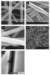
Different morphological structures of fibers can be obtained by altering the electrospinning parameters: (a) cylindrical shape with smooth surface (unpublished original picture by the authors), (b) flat ribbon like morphology (unpublished original picture by the authors), (c) porous (unpublished original picture by the authors), (d) bead-on-string morphology (unpublished original picture by the authors), and (e) core-shell structure (reproduced from open access article [63] under the terms of the Creative Commons Attribution-Non Commercial License.) All these morphologies are obtained through the electrospinning of polylactide.
Figure 3
Figure 3
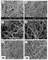
Different arrangements of fiber deposits obtained through electrospinning: (a) random, (b) oriented, and (c) yarn. Unpublished original pictures by the authors. All these structures are obtained through the electrospinning of PLA (original images from the authors).
Figure 4
Figure 4
Fabrication techniques of biomolecule-loaded electrospun fibers.
Figure 5
Figure 5
Flow chart showing biopolymers according to diverse origins and types.
Figure 6
Figure 6
(a)The application of a bacterial cellulose-based scaffold as a biological dressing on burned facial skin and (b) complete epithelialization obtained after two weeks by using bacterial cellulose (BC) as a temporary skin substitute. Reproduced with permission from [103] (License number: 4875380107950).
Figure 7
Figure 7
SEM images of (a) oxidized cellulose fibers, (b) cellulose-chitosan fibers, (c) cellulose-poly (methylmethacrylate) (PMMA) reproduced with permission from [38] (License Number: 4873720243859), (d) poly (2-hydroxy ethyl methacrylate) (pHEMA)-bamboo cellulose nanocomposite fiber reproduced with permission from [108] (License Number: 4873710894891), (e) CA/honey nanofibrous mesh reproduced with permission from [37] (License Number: 4873720705310), (f) Transmission electron microscopy (TEM) image of the ZnO/CA composite fiber reproduced with permission from [113] (License Number: 4873720979763).
Figure 8
Figure 8
SEM images of (a) chitin and (b) deacetylated chitin (chitosan) nanofibrous matrix reproduced with permission from [117] (License Number: 4875380936021), (c) randomly oriented fibrous mesh of chitosan/polyethylene oxide (PEO) (1:1) (d) randomly oriented fibrous mesh of chitosan/PEO/chitin nanocrystals (ChNC) reproduced with permission from [118] (License Number: 4875390863950), (e) chitosan/PEO (90/10) nanofibrous matrix, (f) chitosan/PEO (90/10) nanofibrous matrix blend with 1 wt% Henna extract reproduced with permission from [119] (License Number: 4875401354853).
Figure 9
Figure 9
SEM images of (a) poly(hydroxybutyrate) (PHB)/Collagen (50/50 wt%) electrospun fiber meshes reproduced from open access article [143] distributed under the Creative Commons Attribution License, (b) poly(3-hydroxybutyrate-co-4-hydroxybutyrate) P(3HB)/P(3HO-co-3HD) electrospun fiber meshes functionalized with electrosprayed chitin-lignin/glycyrrhizin acid (CLA) (c) poly (3-hydroxybutyrate-co-3-hydroxyvalerate) (PHBV)/Curcumin (0.5) electrospun fiber meshes and cross-sections of drug-loaded nanofibers and (d) SEM images of L929 fibroblast cells cultured on PHBV/Curcumin (0.5 w/v%) electrospun fiber meshes after incubation for 14 days reproduced from [39] (License Number: 4876940110497).

References

    1. Adeli H., Khorasani M.T., Parvazinia M. Wound dressing based on electrospun PVA/chitosan/starch nanofibrous mats: Fabrication, antibacterial and cytocompatibility evaluation and in vitro healing assay. Int. J. Biol. Macromol. 2019;122:238–254. doi: 10.1016/j.ijbiomac.2018.10.115. - DOI - PubMed
    1. Memic A., Abudula T., Mohammed H.S., Joshi Navare K., Colombani T., Bencherif S.A. Latest Progress in Electrospun Nanofibers for Wound Healing Applications. ACS Appl. Bio. Mater. 2019;2:952–969. doi: 10.1021/acsabm.8b00637. - DOI - PubMed
    1. Augustine R., Rehman S.R.U., Ahmed R., Zahid A.A., Sharifi M., Falahati M., Hasan A. Electrospun chitosan membranes containing bioactive and therapeutic agents for enhanced wound healing. Int. J. Biol. Macromol. 2020;156:153–170. doi: 10.1016/j.ijbiomac.2020.03.207. - DOI - PubMed
    1. Zhu Z., Liu Y., Xue Y., Cheng X., Zhao W., Wang J., He R., Wan Q., Pei X. Tazarotene Released from Aligned Electrospun Membrane Facilitates Cutaneous Wound Healing by Promoting Angiogenesis. ACS Appl. Mater. Interfaces. 2019;11:36141–36153. doi: 10.1021/acsami.9b13271. - DOI - PubMed
    1. Chen Q., Wu J., Liu Y., Li Y., Zhang C., Qi W., Yeung K.W.K., Wong T.M., Zhao X., Pan H. Electrospun chitosan/PVA/bioglass Nanofibrous membrane with spatially designed structure for accelerating chronic wound healing. Mater. Sci. Eng. C Mater. Biol. Appl. 2019;105:110083. doi: 10.1016/j.msec.2019.110083. - DOI - PubMed

LinkOut - more resources