

MiRNA Profiles of Extracellular Vesicles Secreted by Mesenchymal Stromal Cells-Can They Predict Potential Off-Target Effects?
- PMID: 32971982
- PMCID: PMC7565205
- DOI: 10.3390/biom10091353
MiRNA Profiles of Extracellular Vesicles Secreted by Mesenchymal Stromal Cells-Can They Predict Potential Off-Target Effects?
Abstract
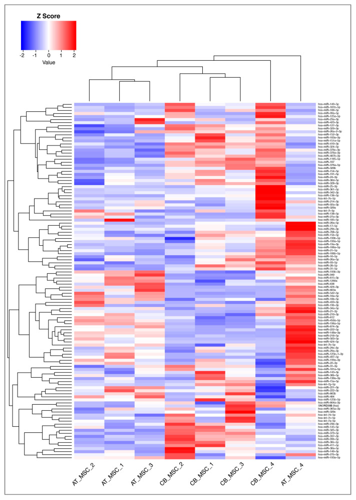
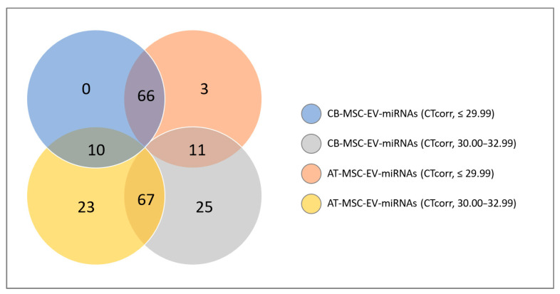
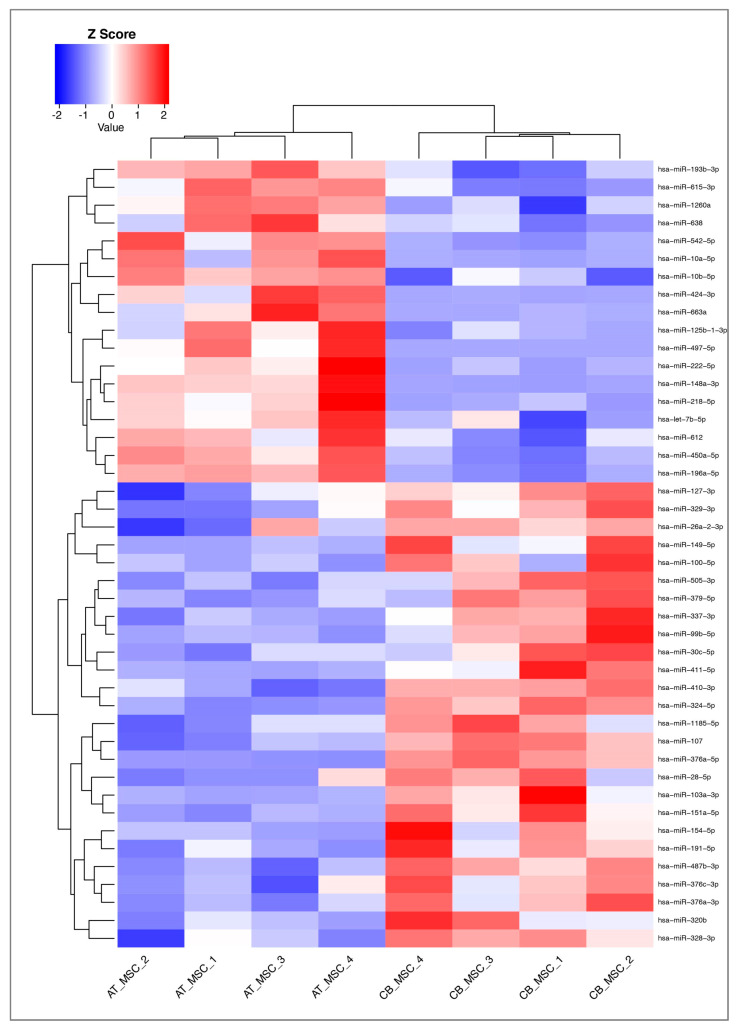
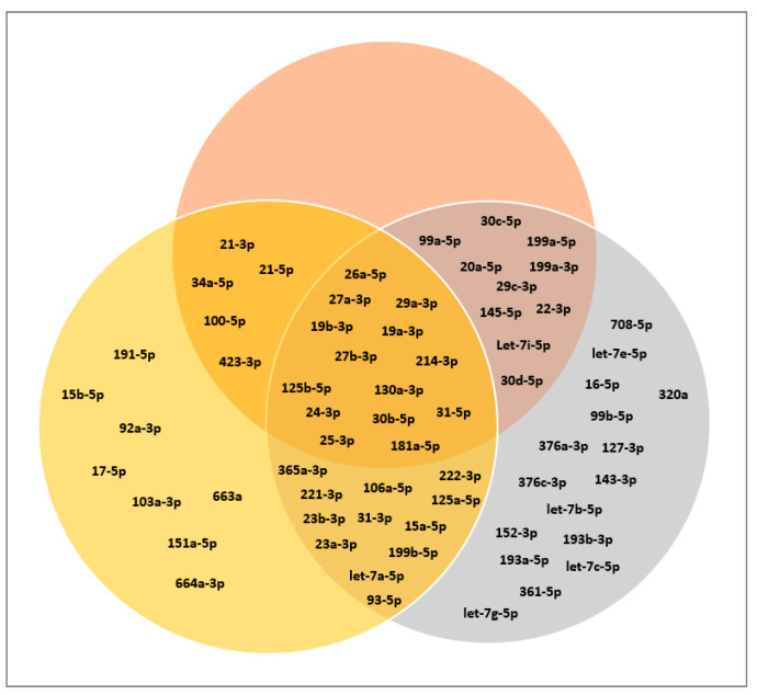
The cardioprotective properties of extracellular vesicles (EVs) derived from mesenchymal stromal cells (MSCs) are currently being investigated in preclinical studies. Although microRNAs (miRNAs) encapsulated in EVs have been identified as one component responsible for the cardioprotective effect of MSCs, their potential off-target effects have not been sufficiently characterized. In the present study, we aimed to investigate the miRNA profile of EVs isolated from MSCs that were derived from cord blood (CB) and adipose tissue (AT). The identified miRNAs were then compared to known targets from the literature to discover possible adverse effects prior to clinical use. Our data show that while many cardioprotective miRNAs such as miR-22-3p, miR-26a-5p, miR-29c-3p, and miR-125b-5p were present in CB- and AT-MSC-derived EVs, a large number of known oncogenic and tumor suppressor miRNAs such as miR-16-5p, miR-23a-3p, and miR-191-5p were also detected. These findings highlight the importance of quality assessment for therapeutically applied EV preparations.
Keywords: adipose tissue; cardioprotection; cord blood; extracellular vesicles; mesenchymal stromal cells; microRNA; oncomiR; tumor suppressor.
Conflict of interest statement
The authors declare no conflict of interest.
Figures
Similar articles
-
Identification of miRNA Reference Genes in Extracellular Vesicles from Adipose Derived Mesenchymal Stem Cells for Studying Osteoarthritis.Int J Mol Sci. 2019 Mar 5;20(5):1108. doi: 10.3390/ijms20051108. Int J Mol Sci. 2019. PMID: 30841483 Free PMC article.
-
Insights into Inflammatory Priming of Adipose-Derived Mesenchymal Stem Cells: Validation of Extracellular Vesicles-Embedded miRNA Reference Genes as A Crucial Step for Donor Selection.Cells. 2019 Apr 23;8(4):369. doi: 10.3390/cells8040369. Cells. 2019. PMID: 31018576 Free PMC article.
-
Mesenchymal Stromal Cell-Derived Extracellular Vesicles Attenuate Dendritic Cell Maturation and Function.Front Immunol. 2018 Nov 9;9:2538. doi: 10.3389/fimmu.2018.02538. eCollection 2018. Front Immunol. 2018. PMID: 30473695 Free PMC article.
-
Therapeutic prospects of MicroRNAs carried by mesenchymal stem cells-derived extracellular vesicles in autoimmune diseases.Life Sci. 2021 Jul 15;277:119458. doi: 10.1016/j.lfs.2021.119458. Epub 2021 Apr 6. Life Sci. 2021. PMID: 33831424 Review.
-
Mesenchymal Stromal Cell-Derived Extracellular Vesicles Regulate the Mitochondrial Metabolism via Transfer of miRNAs.Front Immunol. 2021 Mar 16;12:623973. doi: 10.3389/fimmu.2021.623973. eCollection 2021. Front Immunol. 2021. PMID: 33796099 Free PMC article. Review.
Cited by
-
Extracellular Vesicles of Mesenchymal Stem Cells: Therapeutic Properties Discovered with Extraordinary Success.Biomedicines. 2021 Jun 10;9(6):667. doi: 10.3390/biomedicines9060667. Biomedicines. 2021. PMID: 34200818 Free PMC article. Review.
-
MSC-derived extracellular vesicles: Precision miRNA delivery for overcoming cancer therapy resistance.Regen Ther. 2025 Apr 1;29:303-318. doi: 10.1016/j.reth.2025.03.006. eCollection 2025 Jun. Regen Ther. 2025. PMID: 40237010 Free PMC article. Review.
-
Treatment of Cardiac Fibrosis with Extracellular Vesicles: What Is Missing for Clinical Translation?Int J Mol Sci. 2023 Jun 22;24(13):10480. doi: 10.3390/ijms241310480. Int J Mol Sci. 2023. PMID: 37445658 Free PMC article. Review.
-
Preparation of Recombinant Human Collagen III Protein Hydrogels with Sustained Release of Extracellular Vesicles for Skin Wound Healing.Int J Mol Sci. 2022 Jun 3;23(11):6289. doi: 10.3390/ijms23116289. Int J Mol Sci. 2022. PMID: 35682968 Free PMC article.
-
MSC-EV therapy for bone/cartilage diseases.Bone Rep. 2022 Nov 9;17:101636. doi: 10.1016/j.bonr.2022.101636. eCollection 2022 Dec. Bone Rep. 2022. PMID: 36389627 Free PMC article.
References
-
- van der Spoel T.I., Jansen of Lorkeers S.J., Agostoni P., van Belle E., Gyöngyösi M., Sluijter J.P., Cramer M.J., Doevendans P.A., Chamuleau S.A. Human relevance of pre-clinical studies in stem cell therapy: Systematic review and meta-analysis of large animal models of ischaemic heart disease. Cardiovasc. Res. 2011;91:649–658. doi: 10.1093/cvr/cvr113. - DOI - PubMed
-
- Jansen of Lorkeers S.J., Eding J.E., Vesterinen H.M., van der Spoel T.I., Sena E.S., Duckers H.J., Doevendans P.A., Macleod M.R., Chamuleau S.A. Similar effect of autologous and allogeneic cell therapy for ischemic heart disease: Systematic review and meta-analysis of large animal studies. Circ. Res. 2015;116:80–86. doi: 10.1161/CIRCRESAHA.116.304872. - DOI - PubMed
Publication types
MeSH terms
Substances
Grants and funding
LinkOut - more resources
Full Text Sources
