. 2020 Sep 22;21(18):6974.
doi: 10.3390/ijms21186974.

A Novel Cis-Acting RNA Structural Element Embedded in the Core Coding Region of the Hepatitis C Virus Genome Directs Internal Translation Initiation of the Overlapping Core+1 ORF

Affiliations

A Novel Cis-Acting RNA Structural Element Embedded in the Core Coding Region of the Hepatitis C Virus Genome Directs Internal Translation Initiation of the Overlapping Core+1 ORF

Niki Vassilaki et al. Int J Mol Sci. .

Abstract

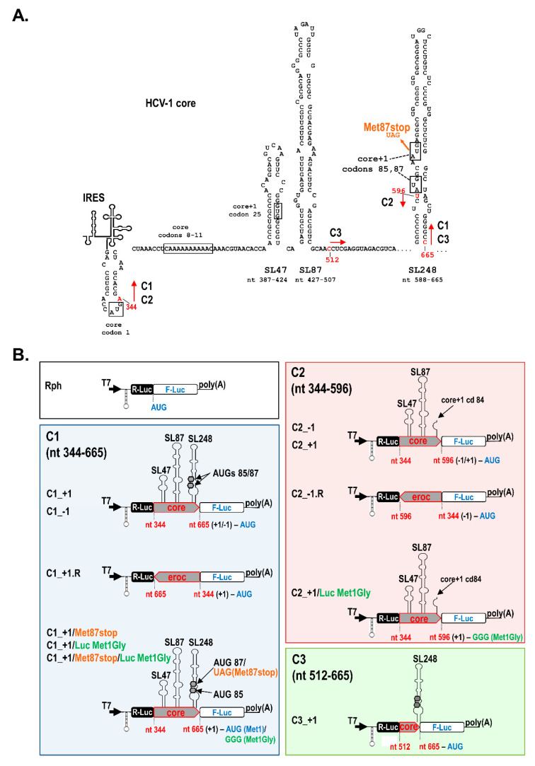
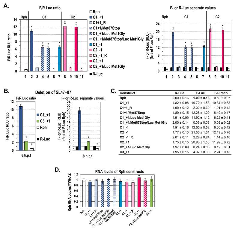
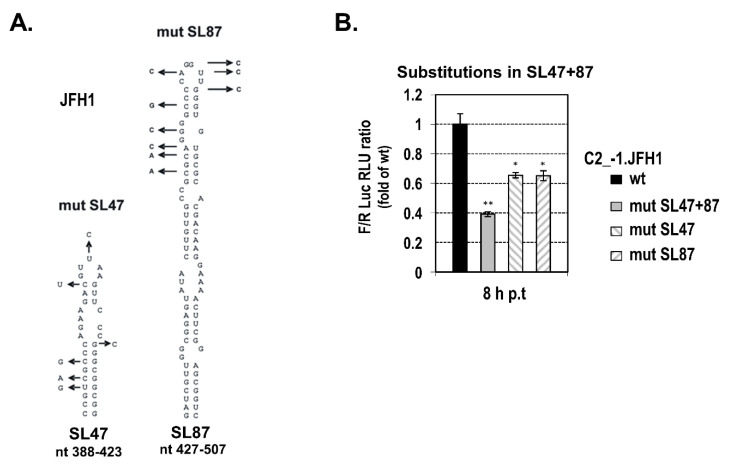
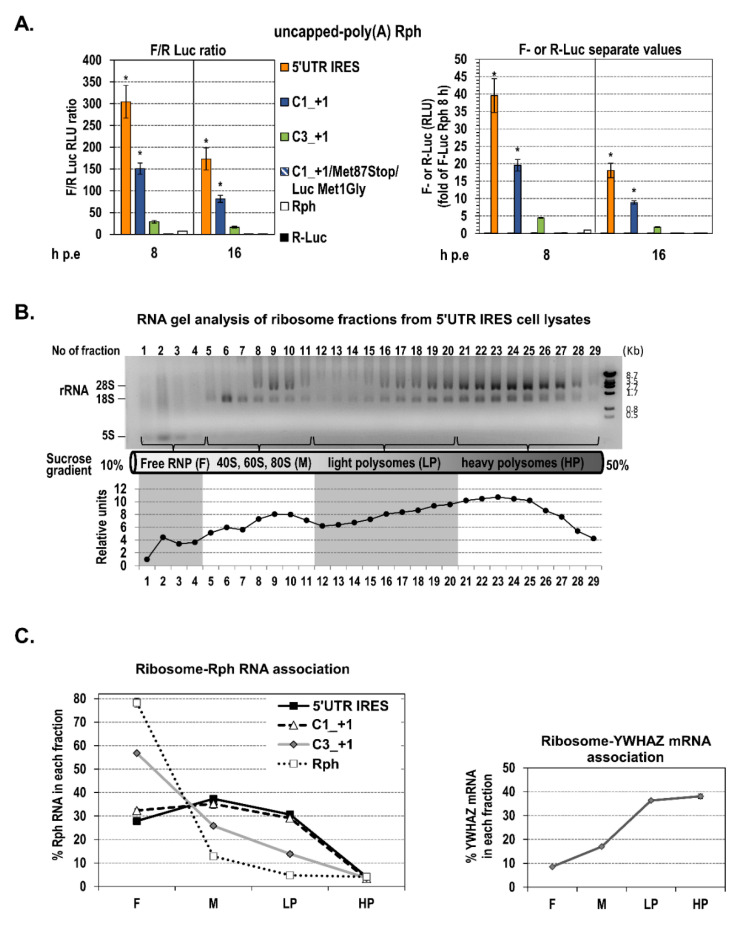
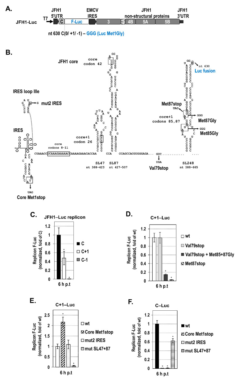
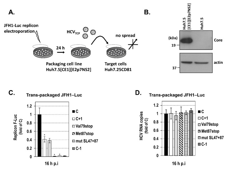
Hepatitis C virus (HCV) genome translation is initiated via an internal ribosome entry site (IRES) embedded in the 5'-untranslated region (5'UTR). We have earlier shown that the conserved RNA stem-loops (SL) SL47 and SL87 of the HCV core-encoding region are important for viral genome translation in cell culture and in vivo. Moreover, we have reported that an open reading frame overlapping the core gene in the +1 frame (core+1 ORF) encodes alternative translation products, including a protein initiated at the internal AUG codons 85/87 of this frame (nt 597-599 and 603-605), downstream of SL87, which is designated core+1/Short (core+1/S). Here, we provide evidence for SL47 and SL87 possessing a novel cis-acting element that directs the internal translation initiation of core+1/S. Firstly, using a bicistronic dual luciferase reporter system and RNA-transfection experiments, we found that nucleotides 344-596 of the HCV genotype-1a and -2a genomes support translation initiation at the core+1 frame AUG codons 85/87, when present in the sense but not the opposite orientation. Secondly, site-directed mutagenesis combined with an analysis of ribosome-HCV RNA association elucidated that SL47 and SL87 are essential for this alternative translation mechanism. Finally, experiments using cells transfected with JFH1 replicons or infected with virus-like particles showed that core+1/S expression is independent from the 5'UTR IRES and does not utilize the polyprotein initiation codon, but it requires intact SL47 and SL87 structures. Thus, SL47 and SL87, apart from their role in viral polyprotein translation, are necessary elements for mediating the internal translation initiation of the alternative core+1/S ORF.

Keywords: RNA translation; core+1 ORF; hepatitis C virus; stem-loops.


Conflict of interest statement

The authors declare no conflict of interest. The funders had no role in the design of the study; in the collection, analyses, or interpretation of data; in the writing of the manuscript, or in the decision to publish the results.

Figures

Figure 1
Figure 1
Schematic representation of the bicistronic plasmids constructed to investigate the mechanism directing core+1/Short (core+1/S) expression. (A) Scheme of HCV-1 (Hepatitis C virus) core coding region, including arrows indicating the start and end positions of the sequences (C1, C2, C3) inserted in the dual luciferase plasmids, relative to the predicted RNA secondary structures SL47, SL87, and SL248. (B) Diagram of the bicistronic constructs used. The parental vector is Renilla-photinus hairpin (Rph) (B top left), which has a stable hairpin (h) loop immediately upstream of the R-Luc gene and carries the Rph cassette under the control of a T7 promoter. Between the R-Luc and Firefly luciferase (F-Luc) genes, the parts of the HCV-1 core nucleotide sequence 344–665 (C1), 344–596 (C2), or 512–665 (C3) were cloned. Core sequences were inserted in the sense 5′-3′ or reverse (R) 3′-5′ orientation, and F-Luc gene was fused in the core+1 or core−1 frame. The Met87stop non-sense mutation at core+1 frame codon 87 (panels a, b for C1 construct) and Luc Met1Gly double substitution converting the F-Luc initiation codon ATG to glycine codon GGG (panel b for C1 and C2 constructs) are also illustrated.
Figure 2
Figure 2
Evidence for a novel cis-acting element within the core coding sequence with capacity for internal translation initiation. (A) The F-Luc to R-Luc values ratio (F/R) or separate values of F-Luc and R-Luc activities detected in Huh7 cells after transfection with in vitro transcribed, capped, and polyA-tailed Rph-derived RNA containing either C1 or C2 core nucleotide sequence, in 5′-3′ or reverse (R) orientation, and the F-Luc gene fused in the core+1 or core−1 frame, in the absence or presence of the mutation Met87stop and/or substitution Luc Met1Gly. Cells were cultured for 8 h post-transfection initiation (h p.t). Values are means ± SD of four independent experiments in triplicate, expressed as relative light unit (RLU) ratio (left) or as separate F-Luc and R-Luc RLUs (right). F-Luc and R-Luc values obtained from cells transfected with the different constructs were expressed relative to the F-Luc value of the negative control empty vector Rph, which was set to one, and their ratios were calculated. Total protein amount was used for normalization. * p < 0.001 cells transfected with C1, C2 constructs vs. Rph empty vector, 5′-3′ constructs vs. R ones, mutated constructs vs. wild-type ones (Student’s t test). (B) Ratio F/R (left) or separate values (right) of F-Luc and R-Luc activities derived from Huh7 cells after transfection with in vitro transcribed, capped, and polyA-tailed C3_+1 Rph RNA as compared to the C1_+1 Rph construct. Cells were lysed 8 h p.t. Mean values were obtained from four independent experiments in triplicate and expressed as RLU ratio (left) or as separate F-Luc and R-Luc RLUs (right). F-Luc and R-Luc values were expressed relative to the F-Luc value derived from cells transfected with the negative control Rph, which was set to one, and their ratios were calculated. * p < 0.001, vs. C1_+1-transfected cells (Student’s t test). (C) Table presenting the data of panel (A) and panel (B) graphs. (D) RNA levels quantified 8 h p.t. of Huh7 cells with in vitro transcribed RNAs from the different Rph constructs, by RT-qPCR analysis of F-Luc gene. The expression of YWHAZ cellular gene was used for normalization. Three independent experiments were performed.
Figure 3
Figure 3
Mutation analysis of SL47 and SL87. (A) Schematic representation of the predicted secondary structures of SL47 and SL87 of JFH1 including substitutions disrupting base-pair interaction (indicated with arrows). The mutations do not affect the amino acid sequence of the core protein. (B) Ratio F/R of F-Luc to R-Luc activity derived from Huh7 cells transfected with a C2_−1.JFH1 Rph RNA, containing either the wild-type C2 core nt fragment 344–596 of JFH1, with the F-Luc gene fused in the core−1 frame (control, set to 1), or a mutated variant carrying the substitutions specified at panel a, within SL47 (C2_−1. JFH1/mut SL47), SL87 (C2_−1. JFH1/mut SL87) or both SL47 and SL87 (C2_−1. JFH1/mut SL47+87). Cells were further cultured after transfection initiation for 8 h. Values are means ± SD of four independent experiments in triplicate, * p < 0.01, ** p < 0.001, vs. control-transfected cells (Student’s t test).
Figure 4
Figure 4
Distribution of the Rph constructs-derived RNAs in the ribosomal fractions of Huh-7 cells. (A) Ratio F/R (left) or separate values (right) of F-Luc and R-Luc activities determined in Huh7 cells after electroporation with in vitro transcribed, uncapped, and polyA-tailed RNA of the Rph-based 5′-untranslated region (5′UTR) internal ribosome entry site (IRES), C1_+1, C3_+1, or C1_+1/Met87stop/Luc Met1Gly constructs or the empty vector Rph, 8 and 16 h post-electroporation (p.e). Values are means ± SD of three independent experiments in triplicate and expressed as RLU ratio (left) or as separate F-Luc and R-Luc RLUs (right). F-Luc and R-Luc values for all constructs were expressed relative to the F-Luc value derived from cells transfected with the negative control Rph at 8 h p.t, which was set to one, and their ratios were calculated. * p < 0.001 vs. C3_+1 and vs. Rph transfected cells (Student’s t test). (B,C) The time-point of 16 h p.e. was selected to examine the loading of ribosomes on the uncapped RNAs of the Rph-based 5′UTR IRES, C1_+1, C3_+1 sequences and the respective empty vector Rph after electroporation in Huh7 cells. Cell lysates were subjected to sucrose density gradient ultracentrifugation. The distribution of ribosomal RNA along the collected fractions was determined by agarose gel electrophoresis (B, top) and quantified by Quantity One software (B, bottom). A representative gel analysis of three independent experiments is shown (of cell lysates from cells transfected with 5′UTR IRES RNA). The positions of fractions with free ribonucleoprotein complexes (F, fractions 1–4), 40S–60S–80S monosomes (M, fractions 5–11), light polysomes (LP, fractions 12–20), and heavy polysomes (HP, fractions 21–29) are depicted with arrows at the bottom of the gel, where a schematic of the 10–50% sucrose gradient is presented. Fractions from the above groups were pooled, total RNA was extracted, and the ribosome–HCV RNA association was analyzed with RT-qPCR (C left). The distribution of the mRNA of the housekeeping gene 14-3-3-zeta polypeptide (YWHAZ) was also analyzed, as a control (C right).
Figure 5
Figure 5
Core+1/S expression in the context of JFH1 replicon in transfected cells. (A) Schematic representation of the bicistronic subgenomic reporter JFH1 replicons (JFH1‒Luc) used. In the replicons, the F-Luc gene was fused to the nucleotides 1–630 of JFH1 strain, in the core (C), core+1 (C+1) or core−1 (C−1) frame. The initiator ATG of Luc has been mutated to GGG (Gly). (B) Schematic representation depicting the position of the mutations inserted in the JFH1‒Luc replicon, relative to the predicted core-region RNA secondary structures: non-sense substitutions at core+1 frame codon 79 abolishing all putative forms of core+1 starting before ATG initiators 85/87 and at codon 87, which in addition abrogates the expression of core+1/S, substitutions converting the core+1 initiation codons 85 and 87 to GGG (glycine), and the non-sense substitution at core codon 1, designed to abrogate polyprotein expression. In addition, the nucleotide sequence of the HCV-1 IRES loop IIIe is shown including the mut2 IRES single substitution, which is known to completely abolish IRES activity [59]. The introduced mutations are indicated with arrows that point to the substituted nucleotides. (C) Open reading frame overlapping the core gene in the +1 frame (Core+1 ORF) expression at the early stages of HCV replication/translation cycle. Huh7-Lunet cells were transfected with the wild-type core (C), core+1 (C+1), or core−1 (C−1) Luc replicons together with an in vitro transcribed, capped, and polyA-tailed R-Luc expressing RNA used to correct for differences in transfection efficiency. F-Luc produced by the replicons was measured at 6 h p.t. and normalized against the renilla luciferase activity derived from the co-transfected R-Luc RNA. Values for the C‒Luc replicon were set to 1 (control). Three independent experiments in triplicate were performed. (D) Core+1 ORF mutation analysis for elucidating the site of core+1/S translation initiation. F-Luc activity in Huh7-Lunet cells that were transfected with the wild-type C+1‒Luc replicon (control, set to 1) or a mutant carrying a non-sense mutation (stop) at core+1 frame codon 79 (Val79stop) or codon 87 (Met87stop), or Val79stop combined with glycine converting mutations at codons 85/87 (Met85+87Gly). Luciferase values were measured and normalized as described in C. Three independent experiments in triplicate were performed. (E,F) IRES mutation analysis for elucidating the mechanism of core+1/S translation. F-Luc activity in Huh7-Lunet cells that were transfected with the wild-type C+1 (E) or C (F) luciferase replicons (control, set to 1) or one of the respective mutants carrying a nucleotide substitution at core codon 1 (core Met1stop) or within IRES (mut2 IRES) or multiple substitutions disrupting RNA stem-loops SL47 and SL87 (mut SL47+87). Luciferase values were measured and normalized as described in C. Three independent experiments in triplicate were performed. Values are means ± SD, * p < 0.001 vs. control-transfected cells (Student’s t test).
Figure 6
Figure 6
Core+1/S expression in the context of JFH1 replicon after infection with trans-complemented (TCP) viral particles. (A) Experimental setup to create trans-complemented HCV particles (HCVTCP) encapsidating subgenomic JFH1-Luc replicons in the stable cell line Huh7.5[CoreE1][E2p7NS2]. (B) Analysis of the CoreE1 transgene expression in electroporated with the JFH1-Luc replicon packaging cells by Western blotting using HCV core- and actin-specific antibodies. (C,D) Infection of Huh7.25-CD81 target cells by HCVTCP viral particles generated upon the electroporation of Huh7.5[CoreE1][E2p7NS2] cells with one of the subgenomic JFH1-Luc replicons C, C+1, C−1, C+1/Val79stop (Val79stop), C+1/Met87stop (Met87stop), or C+1/mut SL47+87 (mut SL47+87). At 16 h post-infection (p.i.), luciferase activity levels derived from the subgenomic replicons were determined, and the intracellular HCV positive strand RNA copies were quantified by RT-qPCR. Three independent experiments were performed. Values are means ± SD, * p < 0.001 cells transfected with C+1 replicon vs. C, and mutated C+1 replicons with Met87stop and mutSL47+87 vs. the wild-type one (Student’s t test).

Similar articles

Cited by

References

    1. Chen S.L., Morgan T.R. The natural history of hepatitis C virus (HCV) infection. Int. J. Med. Sci. 2006;3:47–52. doi: 10.7150/ijms.3.47. - DOI - PMC - PubMed
    1. Wilder J., Patel K. A Review of the Natural History of Chronic Hepatitis C Infection. N. Am. J. Med. Sci. 2014;7:1–7. doi: 10.7156/najms.2014.0701001. - DOI
    1. Hoofnagle J.H. Course and outcome of hepatitis C. Hepatology. 2002;36:S21–S29. doi: 10.1053/jhep.2002.36227. - DOI - PubMed
    1. World Health Organization Hepatitis C. [(accessed on 31 July 2020)]; Available online: https://www.who.int/en/news-room/fact-sheets/detail/hepatitis-c.
    1. Duncan J.D., Urbanowicz R.A., Tarr A.W., Ball J.K. Hepatitis C Virus Vaccine: Challenges and Prospects. Vaccines. 2020;8:90. doi: 10.3390/vaccines8010090. - DOI - PMC - PubMed