Review
. 2020 Sep 22;21(18):6977.
doi: 10.3390/ijms21186977.

How Kinesin-1 Utilize the Energy of Nucleotide: The Conformational Changes and Mechanochemical Coupling in the Unidirectional Motion of Kinesin-1

Affiliations
Review

How Kinesin-1 Utilize the Energy of Nucleotide: The Conformational Changes and Mechanochemical Coupling in the Unidirectional Motion of Kinesin-1

Jingyu Qin et al. Int J Mol Sci. .

Abstract

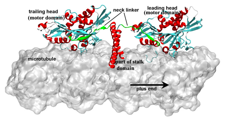
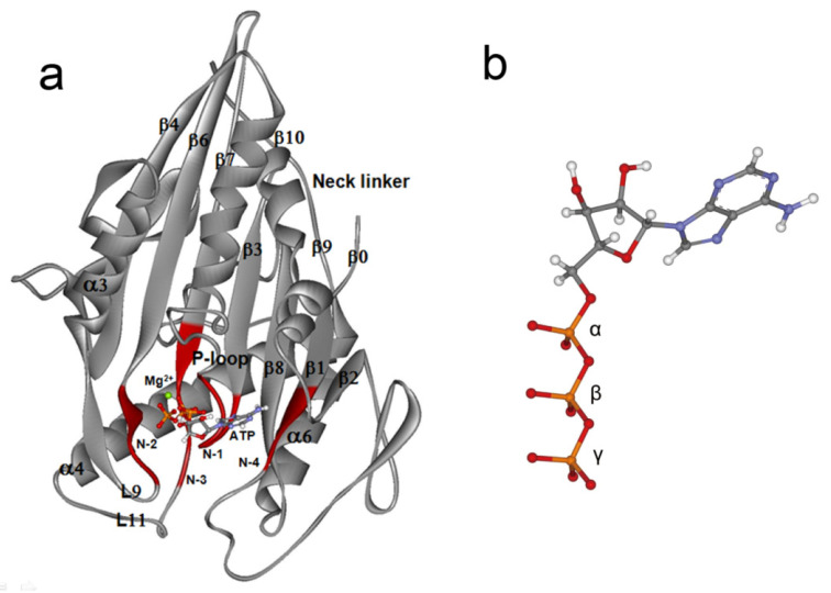
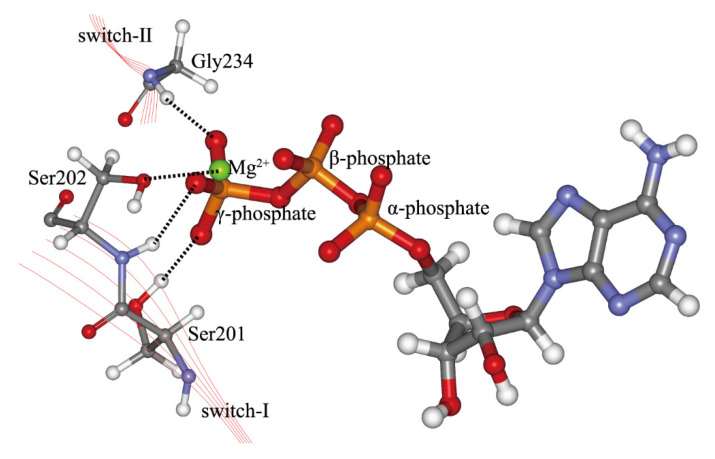
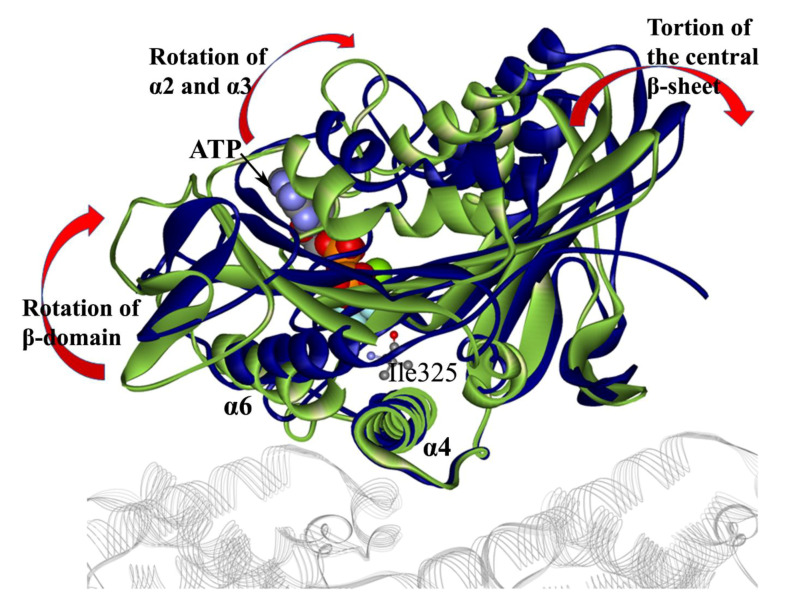
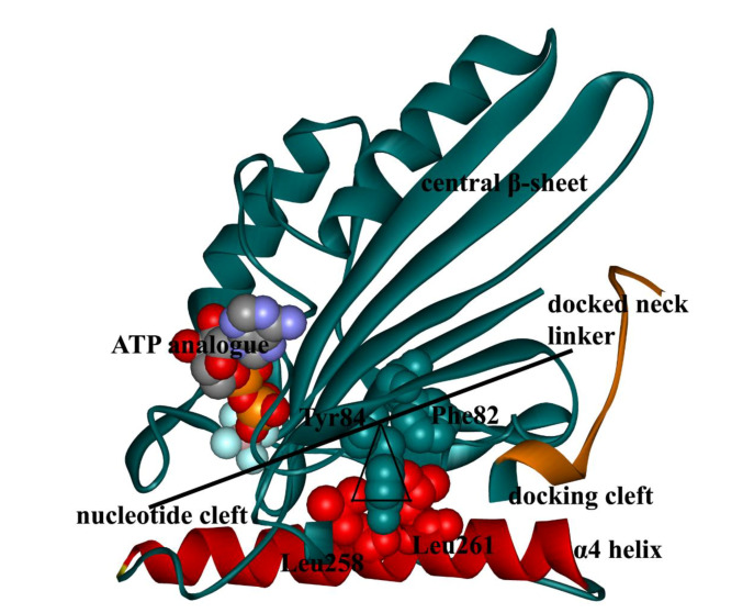
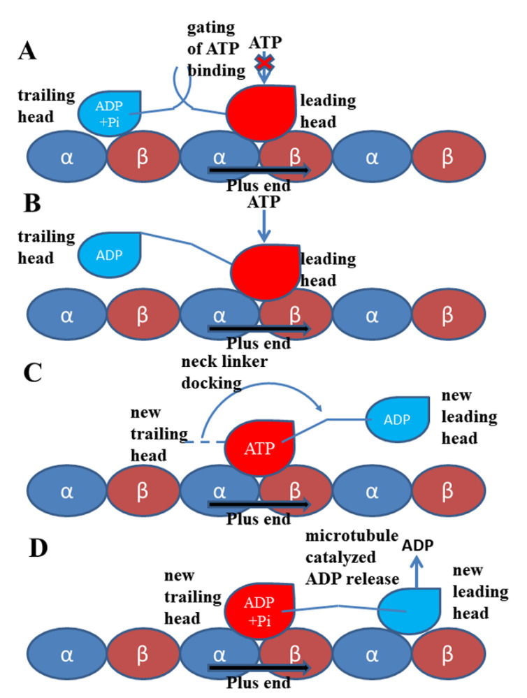
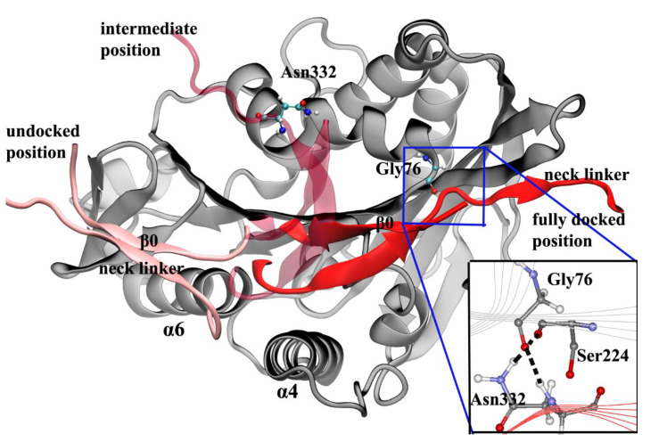
Kinesin-1 is a typical motile molecular motor and the founding member of the kinesin family. The most significant feature in the unidirectional motion of kinesin-1 is its processivity. To realize the fast and processive movement on the microtubule lattice, kinesin-1 efficiently transforms the chemical energy of nucleotide binding and hydrolysis to the energy of mechanical movement. The chemical and mechanical cycle of kinesin-1 are coupled to avoid futile nucleotide hydrolysis. In this paper, the research on the mechanical pathway of energy transition and the regulating mechanism of the mechanochemical cycle of kinesin-1 is reviewed.

Keywords: Kinesin-1; conformational change; mechanochemical coupling; microtubule; neck linker; nucleotide.


Conflict of interest statement

The authors declare no conflict of interest.

Figures

Figure 1
Figure 1
Two motor domains of kinesin-1 bind to the microtubule lattice simultaneously. The neck linkers of the kinesin-1 dimer are colored in green. The leading head is in the nucleotide-free state and has an undocked neck linker, which points to the minus end of the microtubule. The trailing head is in the ADP·Pi/ADP-bound state and has a plus-end pointed neck linker. This figure was produced using Discovery studio 3.5 visualizer.
Figure 2
Figure 2
Structure of kinesin-1 nucleotide-binding pocket and ATP (adenosine triphosphate) molecule. (a) Kinesin-1 motor domain. The four motifs of kinesin-1 nucleotide-binding pocket are highlighted in red. The bound ATP molecule and Mg2+ are shown in the ball and stick mode. (b) Structure of ATP molecule. This figure was produced using Discovery studio 3.5 visualizer.
Figure 3
Figure 3
Interactions of Ser201, Ser202 on switch-I and Gly234 on switch-II with the γ-phosphate of the ATP molecule. The side chain of Ser202 interacts directly with Mg2+. This figure is produced based on the crystal structure 4HNA [61]. The residue numbering of structure 1MKJ [12] is used. This figure was produced by using Discovery studio 3.5 visualizer.
Figure 4
Figure 4
Superposition of the X-ray crystal structures of kinesin-1 and tubulin complex in nucleotide-free (colored in green, PDB ID: 4LNU [62]) and ATP-bound state (colored in blue, PDB ID: 4HNA [61]). The tubulins (represented in the gray line ribbon mode) and α4 helices of the two structures coincide in the superimposed structure. The nucleotide-binding side of the central β-sheet shows a large rotation due to the ATP binding. The microtubule-binding side is relatively stable except for α6, which has a large conformational change. The ATP molecule and the Ile325 (buried in the “docking cleft” when the neck linker is in the docked state) are explicitly shown to depict the position of the ATP-binding site and the “docking cleft” of kinesin-1. This figure was produced by using Discovery studio 3.5 visualizer.
Figure 5
Figure 5
Diagram of the “seesaw” model (PDB ID: 4HNA [61]). The “fulcrum” is composed of Phe82, Tyr84, Leu258 and Leu261. The α4 helix (red) provides the support for the “seesaw”. The 4HNA structure is in the ATP-bound state, with an ATP analogue (ADP-AlF4-) in the nucleotide-binding pocket. In this state, the nucleotide cleft is in the closed state and the docking cleft is in the open state. This figure was produced by using Discovery studio 3.5 visualizer.
Figure 6
Figure 6
Walking and mechanochemical coupling mechanisms of kinesin-1. (A) Both motor domains bind strongly to the microtubule (the same as Figure 1). In this state, the internal strain between the two motor domains and the backward-orientated neck linker of the leading head inhibit the binding of the ATP molecule to this motor domain. (B) Detachment of the trailing head from the microtubule releases the internal strain and the restriction to the orientation of the neck linker of the leading head. The ATP molecule can bind to the leading head. (C) The neck linker docking induced by the ATP binding pulls the trailing head to the next binding site on the microtubule. (D) The ADP-bound state new leading head binds to the microtubule. The ADP molecule releases from this head quickly due to the catalysis of the microtubule.
Figure 7
Figure 7
Diagram of the neck linker docking process. The neck linker in the initial (pink), intermediate (light red), and fully docked positions (red) are shown. In the intermediate position, the distance between the Asn332 and the Gly76 is beyond the force range of a hydrogen bond. The inset is the Asn latch structure in the fully docked state of the neck linker. This figure was produced using VMD (version 1.9.3) [99] and the inset was produced using Discovery studio 3.5 visualizer.

Similar articles

Cited by

References

    1. Lawrence C.J., Kelly Dawe R., Christie K.R., Cleveland D.W., Dawson S.C., Endow S.A., Goldstein L.S.B., Goodson H.V., Hirokawa N., Howard J., et al. A standardized kinesin nomenclature. J. Cell Biol. 2004;167:19–22. doi: 10.1083/jcb.200408113. - DOI - PMC - PubMed
    1. Vale R.D. The molecular motor toolbox for intracellular transport. Cell. 2003;112:467–480. doi: 10.1016/S0092-8674(03)00111-9. - DOI - PubMed
    1. Hirokawa N., Noda Y., Tanaka Y., Niwa S. Kinesin superfamily motor proteins and intracellular transport. Nat. Rev. Mol. Cell Biol. 2009;10:682–696. doi: 10.1038/nrm2774. - DOI - PubMed
    1. Hirokawa N., Tanaka Y. Kinesin superfamily proteins (KIFs): Various functions and their relevance for important phenomena in life and diseases. Exp. Cell Res. 2015;334:16–25. doi: 10.1016/j.yexcr.2015.02.016. - DOI - PubMed
    1. Brady S.T. A novel brain ATPase with properties expected for the fast axonal transport motor. Nature. 1985;317:73–75. doi: 10.1038/317073a0. - DOI - PubMed

LinkOut - more resources