

miR-9-Mediated Inhibition of EFEMP1 Contributes to the Acquisition of Pro-Tumoral Properties in Normal Fibroblasts
- PMID: 32972039
- PMCID: PMC7565260
- DOI: 10.3390/cells9092143
miR-9-Mediated Inhibition of EFEMP1 Contributes to the Acquisition of Pro-Tumoral Properties in Normal Fibroblasts
Abstract
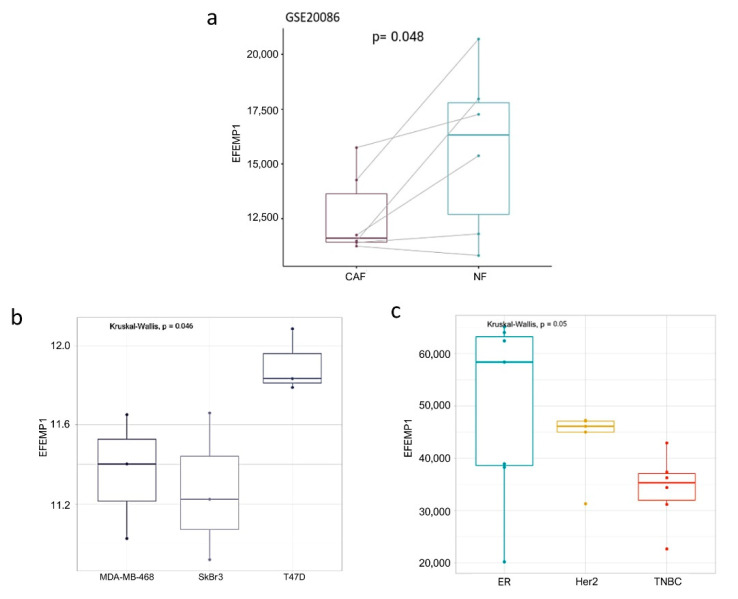
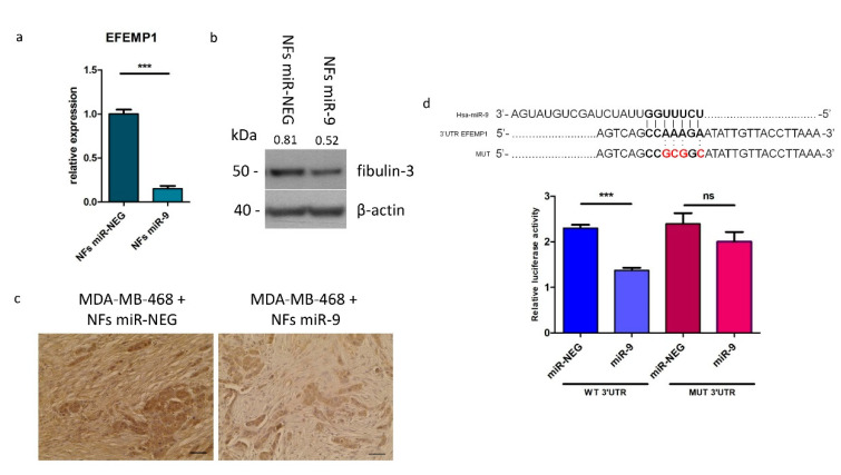
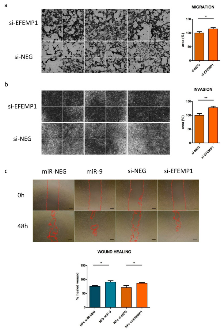
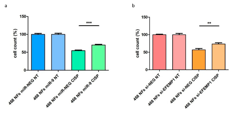
Tumor growth and invasion occurs through a dynamic interaction between cancer and stromal cells, which support an aggressive niche. MicroRNAs are thought to act as tumor messengers to "corrupt" stromal cells. We previously demonstrated that miR-9, a known metastamiR, is released by triple negative breast cancer (TNBC) cells to enhance the transition of normal fibroblasts (NFs) into cancer-associated fibroblast (CAF)-like cells. EGF containing fibulin extracellular matrix protein 1 (EFEMP1), which encodes for the ECM glycoprotein fibulin-3, emerged as a miR-9 putative target upon miRNA's exogenous upmodulation in NFs. Here we explored the impact of EFEMP1 downmodulation on fibroblast's acquisition of CAF-like features, and how this phenotype influences neoplastic cells to gain chemoresistance. Indeed, upon miR-9 overexpression in NFs, EFEMP1 resulted downmodulated, both at RNA and protein levels. The luciferase reporter assay showed that miR-9 directly targets EFEMP1 and its silencing recapitulates miR-9-induced pro-tumoral phenotype in fibroblasts. In particular, EFEMP1 siRNA-transfected (si-EFEMP1) fibroblasts have an increased ability to migrate and invade. Moreover, TNBC cells conditioned with the supernatant of NFs transfected with miR-9 or si-EFEMP1 became more resistant to cisplatin. Overall, our results demonstrate that miR-9/EFEMP1 axis is crucial for the conversion of NFs to CAF-like cells under TNBC signaling.
Keywords: EFEMP1; cancer-associated fibroblasts; chemoresistance; miR-9; miRNA; triple-negative breast cancer; tumor microenvironment.
Conflict of interest statement
The authors declare no conflict of interest. The funders had no role in the design of the study; in the collection, analyses, or interpretation of data; in the writing of the manuscript, or in the decision to publish the results.
Figures
Similar articles
-
Hypoxic cancer-associated fibroblast exosomal circSTAT3 drives triple negative breast cancer stemness via miR-671-5p/NOTCH1 signaling.J Transl Med. 2025 Jul 23;23(1):814. doi: 10.1186/s12967-025-06794-8. J Transl Med. 2025. PMID: 40702555 Free PMC article.
-
Exosome-mediated delivery of miR-9 induces cancer-associated fibroblast-like properties in human breast fibroblasts.Cell Death Dis. 2016 Jul 28;7(7):e2312. doi: 10.1038/cddis.2016.224. Cell Death Dis. 2016. PMID: 27468688 Free PMC article.
-
The stromal loss of miR-4516 promotes the FOSL1-dependent proliferation and malignancy of triple negative breast cancer.Cancer Lett. 2020 Jan 28;469:256-265. doi: 10.1016/j.canlet.2019.10.039. Epub 2019 Oct 28. Cancer Lett. 2020. PMID: 31672492
-
The Role of Cancer-Associated Fibroblasts and Extracellular Vesicles in Tumorigenesis.Int J Mol Sci. 2020 Sep 17;21(18):6837. doi: 10.3390/ijms21186837. Int J Mol Sci. 2020. PMID: 32957712 Free PMC article. Review.
-
miRNAs Delivery for Cancer-associated Fibroblasts' Activation and Drug Resistance in Cancer Microenvironment.Endocr Metab Immune Disord Drug Targets. 2024;24(3):333-347. doi: 10.2174/1871530323666230823094556. Endocr Metab Immune Disord Drug Targets. 2024. PMID: 37612874 Review.
Cited by
-
Secreted Non-Coding RNAs: Functional Impact on the Tumor Microenvironment and Clinical Relevance in Triple-Negative Breast Cancer.Noncoding RNA. 2022 Jan 11;8(1):5. doi: 10.3390/ncrna8010005. Noncoding RNA. 2022. PMID: 35076579 Free PMC article. Review.
-
Fibroblasts as Turned Agents in Cancer Progression.Cancers (Basel). 2023 Mar 28;15(7):2014. doi: 10.3390/cancers15072014. Cancers (Basel). 2023. PMID: 37046676 Free PMC article. Review.
-
Therapeutic Target Discovery for Multiple Myeloma: Identifying Druggable Genes via Mendelian Randomization.Biomedicines. 2025 Apr 5;13(4):885. doi: 10.3390/biomedicines13040885. Biomedicines. 2025. PMID: 40299486 Free PMC article.
-
Unveiling novel biomarkers for platinum chemoresistance in ovarian cancer.Open Med (Wars). 2025 Jan 13;20(1):20241084. doi: 10.1515/med-2024-1084. eCollection 2025. Open Med (Wars). 2025. PMID: 39822989 Free PMC article.
-
The role of cancer-associated fibroblasts in breast cancer metastasis.Front Oncol. 2023 Jul 11;13:1194835. doi: 10.3389/fonc.2023.1194835. eCollection 2023. Front Oncol. 2023. PMID: 37496657 Free PMC article. Review.
References
-
- Dumont N., Liu B., Defilippis R.A., Chang H., Rabban J.T., Karnezis A.N., Tjoe J.A., Marx J., Parvin B., Tlsty T.D. Breast fibroblasts modulate early dissemination, tumorigenesis, and metastasis through alteration of extracellular matrix characteristics. Neoplasia. 2013;15:249–262. doi: 10.1593/neo.121950. - DOI - PMC - PubMed
Publication types
MeSH terms
Substances
LinkOut - more resources
Full Text Sources
Research Materials
Miscellaneous
