

Rule-based automatic diagnosis of thyroid nodules from intraoperative frozen sections using deep learning
- PMID: 32972671
- PMCID: PMC9527708
- DOI: 10.1016/j.artmed.2020.101918
Rule-based automatic diagnosis of thyroid nodules from intraoperative frozen sections using deep learning
Abstract
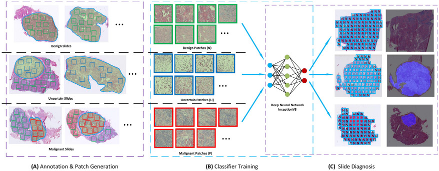
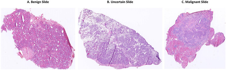
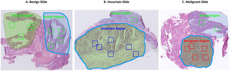
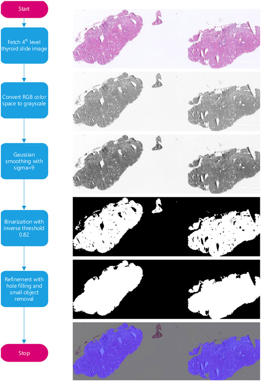
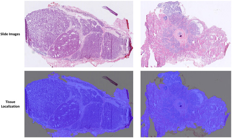
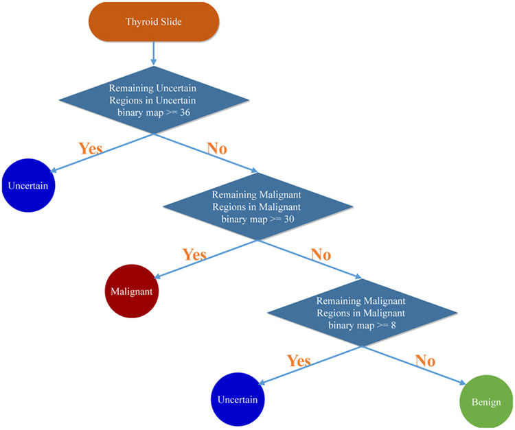
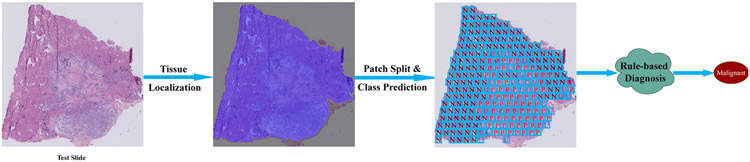
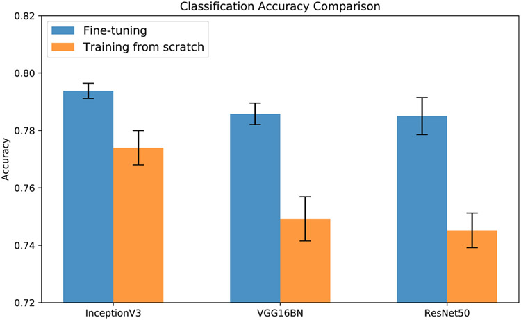
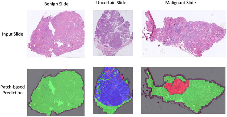
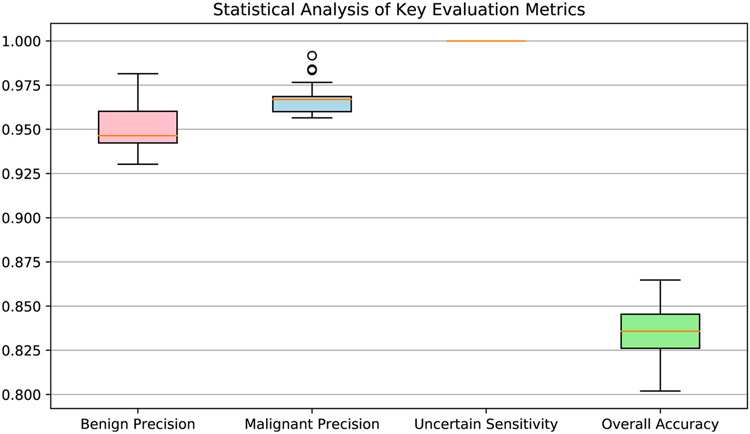
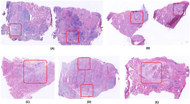
Frozen sections provide a basis for rapid intraoperative diagnosis that can guide surgery, but the diagnoses often challenge pathologists. Here we propose a rule-based system to differentiate thyroid nodules from intraoperative frozen sections using deep learning techniques. The proposed system consists of three components: (1) automatically locating tissue regions in the whole slide images (WSIs), (2) splitting located tissue regions into patches and classifying each patch into predefined categories using convolutional neural networks (CNN), and (3) integrating predictions of all patches to form the final diagnosis with a rule-based system. To be specific, we fine-tune the InceptionV3 model for thyroid patch classification by replacing the last fully connected layer with three outputs representing the patch's probabilities of being benign, uncertain, or malignant. Moreover, we design a rule-based protocol to integrate patches' predictions to form the final diagnosis, which provides interpretability for the proposed system. On 259 testing slides, the system correctly predicts 95.3% (61/64) of benign nodules and 96.7% (148/153) of malignant nodules, and classify 16.2% (42/259) slides as uncertain, including 19 benign and 16 malignant slides, which are a sufficiently small number to be manually examined by pathologists or fully processed through permanent sections. Besides, the system allows the localization of suspicious regions along with the diagnosis. A typical whole slide image, with 80, 000 × 60, 000 pixels, can be diagnosed within 1 min, thus satisfying the time requirement for intraoperative diagnosis. To the best of our knowledge, this is the first study to apply deep learning to diagnose thyroid nodules from intraoperative frozen sections. The code is released at https://github.com/PingjunChen/ThyroidRule.
Keywords: Deep learning; Frozen section; Rule-based protocol; Thyroid nodule; Whole slide image.
Copyright © 2020 Elsevier B.V. All rights reserved.
Conflict of interest statement
Conflict of interests
The authors declare no conflicts of interest.
Figures
Similar articles
-
Pathology diagnosis of intraoperative frozen thyroid lesions assisted by deep learning.BMC Cancer. 2024 Aug 29;24(1):1069. doi: 10.1186/s12885-024-12849-8. BMC Cancer. 2024. PMID: 39210289 Free PMC article.
-
Interactive thyroid whole slide image diagnostic system using deep representation.Comput Methods Programs Biomed. 2020 Oct;195:105630. doi: 10.1016/j.cmpb.2020.105630. Epub 2020 Jun 27. Comput Methods Programs Biomed. 2020. PMID: 32634647 Free PMC article.
-
A study of machine learning models for rapid intraoperative diagnosis of thyroid nodules for clinical practice in China.Cancer Med. 2024 Feb;13(3):e6854. doi: 10.1002/cam4.6854. Epub 2024 Jan 8. Cancer Med. 2024. PMID: 38189547 Free PMC article.
-
Fast and accurate lung cancer subtype classication and localization based on Intraoperative frozen sections of lung adenocarcinoma.Biomed Phys Eng Express. 2025 Jun 16;11(4). doi: 10.1088/2057-1976/ade157. Biomed Phys Eng Express. 2025. PMID: 40472860
-
Thyroid nodules: rational management.World J Surg. 2000 Aug;24(8):934-41. doi: 10.1007/s002680010175. World J Surg. 2000. PMID: 10865037 Review.
Cited by
-
Trends in AI-powered Classification of Thyroid Neoplasms Based on Histopathology Images - a Systematic Review.Acta Inform Med. 2023;31(4):280-286. doi: 10.5455/aim.2023.31.280-286. Acta Inform Med. 2023. PMID: 38379694 Free PMC article.
-
Pathology diagnosis of intraoperative frozen thyroid lesions assisted by deep learning.BMC Cancer. 2024 Aug 29;24(1):1069. doi: 10.1186/s12885-024-12849-8. BMC Cancer. 2024. PMID: 39210289 Free PMC article.
-
Pathomic Features Reveal Immune and Molecular Evolution From Lung Preneoplasia to Invasive Adenocarcinoma.Mod Pathol. 2023 Dec;36(12):100326. doi: 10.1016/j.modpat.2023.100326. Epub 2023 Sep 9. Mod Pathol. 2023. PMID: 37678674 Free PMC article.
-
CellSpatialGraph: Integrate hierarchical phenotyping and graph modeling to characterize spatial architecture in tumor microenvironment on digital pathology.Softw Impacts. 2021 Nov;10:100156. doi: 10.1016/j.simpa.2021.100156. Epub 2021 Oct 9. Softw Impacts. 2021. PMID: 36203948 Free PMC article.
-
The Use of Artificial Intelligence in the Diagnosis and Classification of Thyroid Nodules: An Update.Cancers (Basel). 2023 Jan 24;15(3):708. doi: 10.3390/cancers15030708. Cancers (Basel). 2023. PMID: 36765671 Free PMC article. Review.
References
-
- Novis DA, Gephardt GN, Zarbo RJ. Interinstitutional comparison of frozen section consultation in small hospitals: a college of American pathologists Q-probes study of 18532 frozen section consultation diagnoses in 233 small hospitals. Arch Pathol Lab Med 1996;120(12):1087. - PubMed
-
- Collins KA. The future of the forensic pathology workforce. Acad Forens Pathol 2015;5(4):526–33. 10.23907/2015.058. - DOI
-
- Benediktsson H, Whitelaw J, Roy I. Pathology services in developing countries: a challenge. Arch Pathol Lab Med 2007;131(11):1636–9. - PubMed
MeSH terms
Grants and funding
LinkOut - more resources
Full Text Sources
