

Are Gadolinium-Enhanced MR Sequences Needed in Simultaneous 18F-FDG-PET/MRI for Tumor Delineation in Head and Neck Cancer?
- PMID: 32972956
- PMCID: PMC7661078
- DOI: 10.3174/ajnr.A6764
Are Gadolinium-Enhanced MR Sequences Needed in Simultaneous 18F-FDG-PET/MRI for Tumor Delineation in Head and Neck Cancer?
Abstract
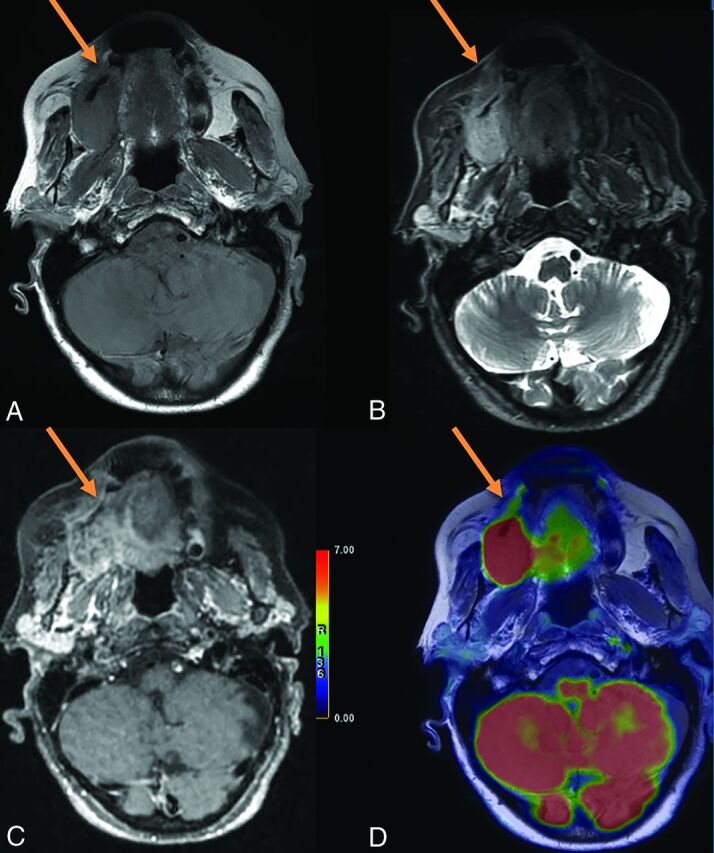
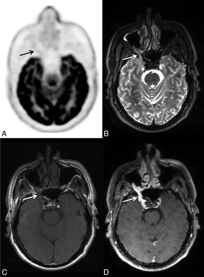
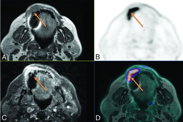
Background and purpose: PET/MRI with 18F-FDG has demonstrated the advantages of simultaneous PET and MR imaging in head and neck cancer imaging, MRI allowing excellent soft-tissue contrast, while PET provides metabolic information. The aim of this study was to evaluate the added value of gadolinium contrast-enhanced sequences in the tumor delineation of head and neck cancers on 18F-FDG-PET/MR imaging.
Materials and methods: Consecutive patients who underwent simultaneous head and neck 18F-FDG-PET/MR imaging staging or restaging followed by surgery were retrospectively included. Local tumor invasion and lymph node extension were assessed in 45 head and neck anatomic regions using 18F-FDG-PET/MR imaging by 2 rater groups (each one including a radiologist and a nuclear medicine physician). Two reading sessions were performed, one without contrast-enhanced sequences (using only T1WI, T2WI, and PET images) and a second with additional T1WI postcontrast sequences. The results were compared with the detailed histopathologic analysis, used as reference standard. The κ concordance coefficient between the reading sessions and sensitivity and specificity for each region were calculated.
Results: Thirty patients were included. There was excellent agreement between the contrast-free and postgadolinium reading sessions in delineating precise tumor extension in the 45 anatomic regions studied (Cohen κ = 0.96, 95% CI = [0.94-0.97], P < .001). The diagnostic accuracy did not differ between contrast-free and postgadolinium reading sessions, being 0.97 for both groups and both reading sessions. For the 2 rater groups, there was good sensitivity for both contrast-free (0.83 and 0.85) and postgadolinium reading sessions (0.88 and 0.90, respectively). Moreover, there was excellent specificity (0.98) for both groups and reading sessions.
Conclusions: Gadolinium chelate contrast administration showed no added value for accurate characterization of head and neck primary tumor extension and could possibly be avoided in the PET/MR imaging head and neck workflow.
© 2020 by American Journal of Neuroradiology.
Figures
Similar articles
-
Contrast-enhanced PET/MR imaging versus contrast-enhanced PET/CT in head and neck cancer: how much MR information is needed?J Nucl Med. 2014 Apr;55(4):551-8. doi: 10.2967/jnumed.113.125443. Epub 2014 Feb 3. J Nucl Med. 2014. PMID: 24491410
-
Qualitative and quantitative performance of ¹⁸F-FDG-PET/MRI versus ¹⁸F-FDG-PET/CT in patients with head and neck cancer.AJNR Am J Neuroradiol. 2014 Oct;35(10):1970-5. doi: 10.3174/ajnr.A3993. Epub 2014 Jun 12. AJNR Am J Neuroradiol. 2014. PMID: 24924545 Free PMC article.
-
[18F]FDG PET/MRI versus contrast-enhanced MRI in detecting regional HNSCC metastases.Ann Nucl Med. 2021 Feb;35(2):260-269. doi: 10.1007/s12149-020-01565-5. Epub 2021 Jan 17. Ann Nucl Med. 2021. PMID: 33454923
-
(18)F-Fluorodeoxyglucose PET/MR Imaging in Head and Neck Cancer.PET Clin. 2016 Oct;11(4):375-86. doi: 10.1016/j.cpet.2016.05.002. Epub 2016 Jul 7. PET Clin. 2016. PMID: 27593244 Review.
-
Diagnostic accuracy of F-18 FDG PET or PET/CT for detection of lymph node metastasis in clinically node negative head and neck cancer patients; A systematic review and meta-analysis.Am J Otolaryngol. 2019 Mar-Apr;40(2):297-305. doi: 10.1016/j.amjoto.2018.10.013. Epub 2018 Oct 23. Am J Otolaryngol. 2019. PMID: 30473166
Cited by
-
PET/MR versus PET/CT for locoregional staging of oropharyngeal squamous cell cancer.Acta Radiol. 2023 May;64(5):1865-1872. doi: 10.1177/02841851221140668. Epub 2022 Dec 4. Acta Radiol. 2023. PMID: 36464816 Free PMC article.
-
Quantitative MRI to Characterize Hypoxic Tumors in Comparison to FMISO PET/CT for Radiotherapy in Oropharynx Cancers.Cancers (Basel). 2023 Mar 22;15(6):1918. doi: 10.3390/cancers15061918. Cancers (Basel). 2023. PMID: 36980806 Free PMC article.
-
PET/MR Imaging in Evaluating Treatment Failure of Head and Neck Malignancies: A Neck Imaging Reporting and Data System-Based Study.AJNR Am J Neuroradiol. 2022 Mar;43(3):435-441. doi: 10.3174/ajnr.A7427. Epub 2022 Feb 17. AJNR Am J Neuroradiol. 2022. PMID: 35177543 Free PMC article.
References
MeSH terms
Substances
LinkOut - more resources
Full Text Sources
Medical