Review
. 2020 Sep 24;11(9):799.
doi: 10.1038/s41419-020-02995-9.

COVID19: an announced pandemic

Affiliations
Review

COVID19: an announced pandemic

Sara Platto et al. Cell Death Dis. .

Abstract

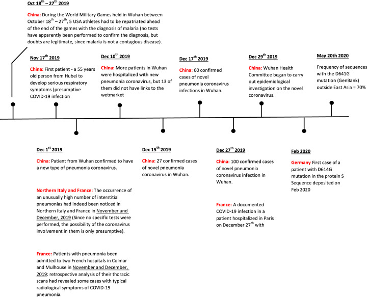
A severe upper respiratory tract syndrome caused by the new coronavirus has now spread to the entire world as a highly contagious pandemic. The large scale explosion of the disease is conventionally traced back to January of this year in the Chinese province of Hubei, the wet markets of the principal city of Wuhan being assumed to have been the specific causative locus of the sudden explosion of the infection. A number of findings that are now coming to light show that this interpretation of the origin and history of the pandemic is overly simplified. A number of variants of the coronavirus would in principle have had the ability to initiate the pandemic well before January of this year. However, even if the COVID-19 had become, so to say, ready, conditions in the local environment would have had to prevail to induce the loss of the biodiversity's "dilution effect" that kept the virus under control, favoring its spillover from its bat reservoir to the human target. In the absence of these appropriate conditions only abortive attempts to initiate the pandemic could possibly occur: a number of them did indeed occur in China, and probably elsewhere as well. These conditions were unfortunately present at the wet marked in Wuhan at the end of last year.


Conflict of interest statement

The authors declare that they have no conflict of interest.

Figures

Fig. 1
Fig. 1. The coronavirus with the Spike trimeric glycoprotein projections from the surface.
a The schematic portion of the Figure (lower left) shows the structural E and M structural proteins of the membrane, the protruding trimeric structural protein, the structural protein N that binds the RN, and the RNA genome. b the 3-D structure of the S-protein with the receptor binding domain (RBD) with the domain that binds the ACE 2 receptor.
Fig. 2
Fig. 2. Philogenetic relationships in the Coronaviridae subfamily from the Coronavirinae family.
The viruses in the subfamily group into four genera: Alphacoronavirus, Betacoronavirus, Gammacoronavirus, and Deltacoronavirus, based on their phylogenic relationshios and genomic structures; the alphacoronaviruses and betacoronaviruses infect only mammals and usually cause respiratory syndromes in humans, the gammacoronaviruses infect avian species and the deltacoronaviruses infect both birds and mammals. The highly infective viruses SARS-CoV and MERS-CoV are betacoronaviruses, the COVID-19 virus is not listed among the betacoronaviruses because it was not yet known and classified in early 2019. Other details are found in Cui et al. 2019 (Figure modified from Cui et al.).
Fig. 3
Fig. 3. A representation of the structure and domain organization of the spike glycoprotein of the coronavirus, which is formed by the two subunits S1 and S2.
At the N-terminus of subunit S1 a signal peptide permits the insertion of the nascent protein into the secretory pathway of the host cell. The S1 subunit contains the receptor binding domain (RBD) that attaches the S protein to the ACE2 receptor of the host cell. The S2 subunit contains the membrane fusion complex (fusion peptide, heptad repeats HR 1 and HR2), anchors the S2 subunits to the viral membrane with its transmembrane domain, and interacts with the viral ribonucleoprotein complex through its endodomain. (modified from Hoffmann et al.).
Fig. 4
Fig. 4. Invasion of the target cell (a pulmonary alveolar cell) by the virus.
Liberation of the viral RNA genomei, its translational activity that produces a number of proteins including interleukin 6, and replicates itself. Other details in the text. (modified from the Web).
Fig. 5
Fig. 5. The proteolytic cleavage site in the S-protein of the SARS-CoV and SARS-CoV-2 viruses.
The protease recognition site (the arginines, R) is different in the two viruses. The arrows indicate the cleavage sites (modified from Hoffmann et al.).
Fig. 6
Fig. 6. 3-D structure protein S of the COVID-19.
a The position of aspartate 614 is conserved among species, until the D-G mutation occurred in the virus variant now dominant in the European and American countries of the pandemic occurred. The D614G mutation generates an additional elastase proteolytic cleavage site that enhances viral membrane fusion next to the S1–S2 junction site (685) (modidfied from Bhattacharyya et al.). b Frequency of the D614/G mutation in the S-protein of COVID-19 in the S-protein sequences available in the GenBank from January to May, 2020. (modified from Zhang et al.).
Fig. 7
Fig. 7
Timeline of the events in the 2020 COVID-19 pandemic.

Similar articles

Cited by

References

    1. Barrett R, Kuzawa CW, McDade T, Armelagos GJ. Emerging and re-emerging infectious diseases: the third epidemiologic transition. Annu. Rev. Anthropol. 1998;27:247–271. doi: 10.1146/annurev.anthro.27.1.247. - DOI
    1. McMichael AJ. Human culture, ecological change, and infectious disease: are we experiencing history’s fourth great transition? Ecosyst. Health. 2001;7:107–115. doi: 10.1046/j.1526-0992.2001.007002107.x. - DOI
    1. Horby, P. W., Hoa, N. ., Pfeiffer, D. U. & Wertheim, H. F. L. Drivers of emerging zoonotic infectious diseases. Confronting Emerging Zoonoses (eds Yamada, A., Kahn, L., Kaplan, B., Monath, T., Woodall, J. & Conti, L.) (Springer Press, Tokyo, 2014).
    1. Wilcox BA, Gubler DJ. Disease ecology and the global emergence of zoonotic pathogens. Environ. Health Prev. Med. 2005;10:263–272. doi: 10.1007/BF02897701. - DOI - PMC - PubMed
    1. Jones KE, et al. Global trends in emerging infectious diseases. Nature. 2008;451:990–994. doi: 10.1038/nature06536. - DOI - PMC - PubMed

MeSH terms