. 2020 Sep 24;10(1):15604.
doi: 10.1038/s41598-020-72450-4.

Aldosterone up-regulates voltage-gated potassium currents and NKCC1 protein membrane fractions

Affiliations

Aldosterone up-regulates voltage-gated potassium currents and NKCC1 protein membrane fractions

Parveen Bazard et al. Sci Rep. .

Abstract

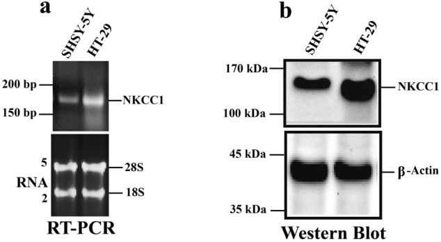
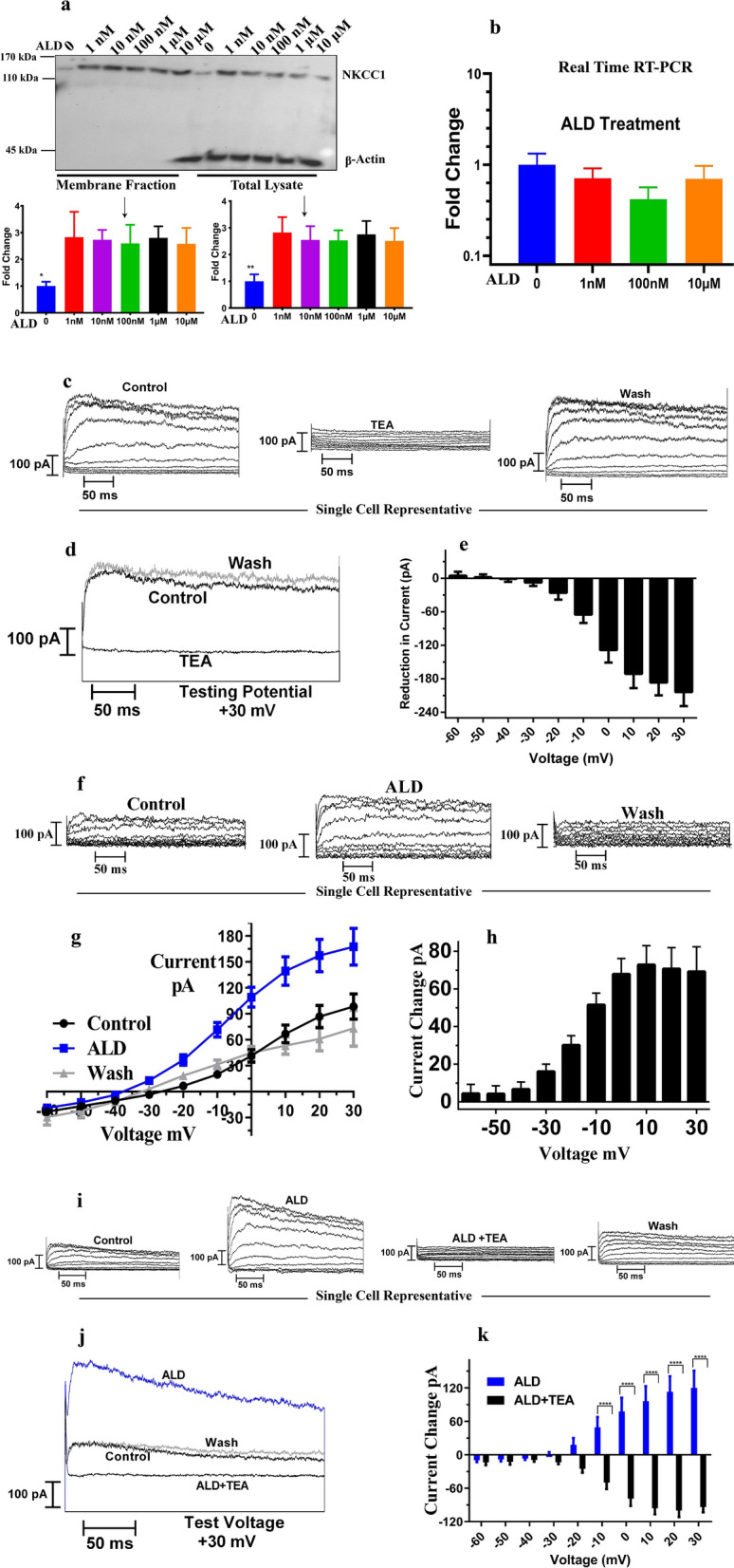
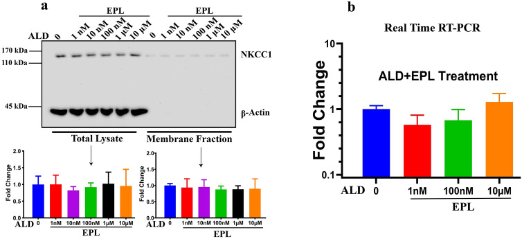
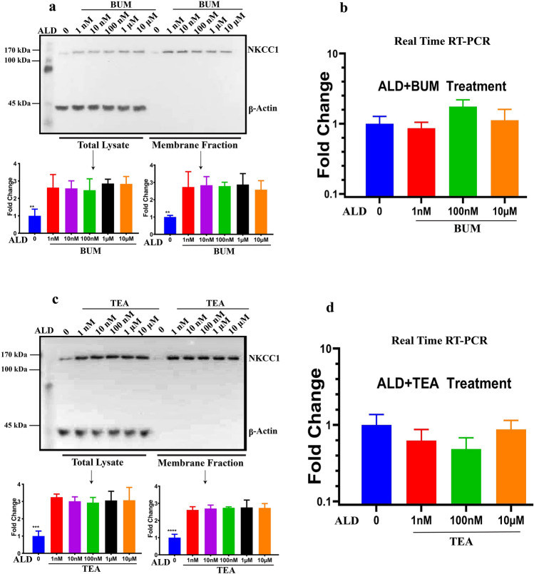
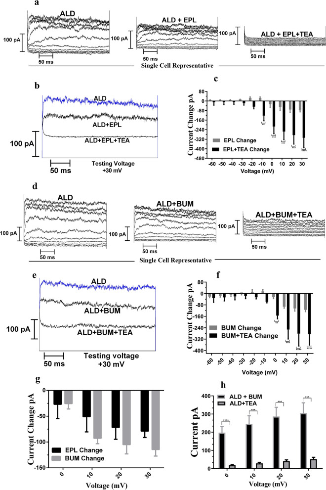
Na+-K+-2Cl- Cotransporter (NKCC1) is a protein that aids in the active transport of sodium, potassium, and chloride ions across cell membranes. It has been shown that long-term systemic treatment with aldosterone (ALD) can enhance NKCC1 protein expression and activity in the aging cochlea resulting in improved hearing. In the present work, we used a cell line with confirmed NKCC1 expression to demonstrate that in vitro application of ALD increased outward voltage-gated potassium currents significantly, and simultaneously upregulated whole lysate and membrane portion NKCC1 protein expression. These ALD-induced changes were blocked by applying the mineralocorticoid receptor antagonist eplerenone. However, application of the NKCC1 inhibitor bumetanide or the potassium channel antagonist Tetraethyl ammonium had no effect. In addition, NKKC1 mRNA levels remained stable, indicating that ALD modulates NKCC1 protein expression via the activation of mineralocorticoid receptors and post-transcriptional modifications. Further, in vitro electrophysiology experiments, with ALD in the presence of NKCC1, K+ channel and mineralocorticoid receptor inhibitors, revealed interactions between NKCC1 and outward K+ channels, mediated by a mineralocorticoid receptor-ALD complex. These results provide evidence of the therapeutic potential of ALD for the prevention/treatment of inner ear disorders such as age-related hearing loss.


Conflict of interest statement

Drs. Ding, Frisina, Zhu and Walton hold a patent (not yet licensed) on the use of aldosterone and anti-inflammatories to treat age-related hearing loss. There are no other actual or perceived conflicts for the authors of this manuscript regarding funding source agencies, NIH or others, for the research reported in this manuscript.

Figures

Figure 1
Figure 1
NKCC1 genes and proteins are expressed in SH-SY5Y neural cells. Gene and protein expressions were analyzed using RT-PCR and western blotting techniques, respectively. (a) RNA was isolated from SH-SY5Y cells and was reverse transcribed, then the cDNA from RT was used for real time PCR (qPCR) to quantify RNA levels. Single band on the right side is HT-29 control cells in both panels a and b. NKCC1 gene expression was detected between 150–200 bp size, for SH-SY5Y cells (a, Top). The bottom lanes show that total RNA was run and the clear 18S and 28S ribosomal RNA bands show that the RNA was intact with no degradation. Total RNA was also used as the loading control for both SH-SY5Y cells and HT-29 Cells and for the evaluation of NKCC1 gene expression difference between SH-SY5Y and HT-29 cell lines (b) Cell lysate was collected in RIPA buffer and analyzed using western blots for NKCC1 protein expression. Data indicate that both SH-SY5Y and HT-29, control cells show NKCC1 expression. For western blot experiments, both proteins of interest, NKCC1 and the loading control protein, β-Actin, were probed on the same PVDF membrane i.e., same membrane were incubated with NKCC1 and β-Actin antibodies. Similarly, for semi-quantitative RT-PCR, both the gene of interest (NKCC1) and reference gene (β-Actin) were probed together using respective primers and analyzed on the same agarose gel.
Figure 2
Figure 2
Aldosterone induces increases in NKCC1 protein expression levels and outward potassium current amplitudes. (a) SH-SY5Y cells were treated with aldosterone (ALD) at concentrations from 1 nM to 10 μM for 24 h. Cell lysates were performed in PBS buffer and membrane fractions were separated using the cell signaling kit. Western blot analysis probed with NKCC1 and β-actin antibodies for both total lysate and membrane fractions. β-actin was used as the loading control and equal amounts of lysate protein were used for the isolation of cell membrane proteins (lysate protein concentration was measured by spectrophotometer). For both total lysate and membrane fractions, NKCC1 protein expression was upregulated following application of ALD as compared to control samples (no ALD treatment). There was no β-actin present in the membrane fractions as it is a structural protein. Data are means ± SD from 3 independent experiments. (b) NKCC1 mRNA was not regulated by ALD treatment. Cell lysates performed in RLT buffer and total RNA was isolated using the Qiagen kit. Subsequently, real-time RT-PCR technique was carried out. There was no significant difference between the control (no ALD) and treated samples (n = 3). All western blot data were taken from the same gel. (c) Whole cell voltage-clamp recordings of a representative SH-SY5Y show outward currents elicited by 300 ms voltage steps. Voltage steps from -60 mV to + 30 mV, in 10 mV step increments were acquired at a holding potential was −90 mV. There was a reversible decrease in K+ currents due to application of tetraethyl ammonium – TEA (1 mM), confirming the presence of K+ channels in SH-SY5Y cells. (d) For a representative cell, current traces at + 30 mV voltage step shows the effects of TEA on K + currents. (e) Similarly, mean reduction in currents with respect to control (n = 6) was shown when cells were perfused with 1 mM TEA. (f) There was a reversible increase in potassium currents when 1 μM ALD was applied as compared to no ALD and currents decreased to the control values when ALD was washed out. (g) Mean I–V functions are plotted for control (n = 17 cells) and after ALD application (n = 5 for wash) were shown as mean current–voltage relations. (h) The change in outward currents with respect to control conditions following ALD application were greater for more positive holding potentials. (i) To further confirm the increase in K+ due to ALD application, ALD was perfused in combination with the K+ blocker, TEA. Currents traces of a representative cell were shown as outward currents in response to test potentials −60 mV to + 30 mV, in 10 mV increments with holding potential −90 mV. There was an increase in potassium currents due to ALD, followed by decrease in currents when TEA was added to the bath. Currents return to control values during wash out. (j, k) Current traces at + 30 mV testing voltage and mean change with respect to control values in currents confirm the same tendency (n = 5). Statistical Significance: *P < 0.05, ** P < 0.01, ***P < 0.001, ****P < 0.0001.
Figure 3
Figure 3
NKCC1 protein levels were increased by upregulation of mineralocorticoid receptors. (a) Cells were treated with eplerenone (EPL—20 µM), a specific mineralocorticoid receptor inhibitor. Cell lysates were collected in PBS buffer and analyzed for total lysate and membrane fraction for NKCC1 protein expressions using western blot techniques. Equal amounts of lysate protein were used for the isolation of cell membrane proteins (according to the lysate protein concentration measured by spectrophotometer). β-actin was used to achieve equal loading for total lysate, but β-actin is not found in membrane fractions. The blue bar is the control, untreated cells and remaining are various colors with concentration of ALD from 1 nM to 10 µM along with EPL. a) There was no difference between control and treated samples (ALD + EPL) i.e., ALD did not upregulate NKCC1 protein expression in the presence of inhibitor. Bar graphs show mean ± SD from 3 independent runs. (b) Similar to protein expression, there was also no difference in gene expression for NKCC1 following ALD treatment in the presence of eplerenone from 1 nM to 10 µM. Bar graph data are presented as mean ± SEM for three independent runs.
Figure 4
Figure 4
The NKCC1 inhibitor bumetanide, and potassium channel blocker TEA, did not alter ALD mediated changes in NKCC1 protein expression via post-transcriptional modification. (a) Cells were treated with ALD in presence bumetanide (BUM—10 μM), a specific NKCC1 activity blocker, with blue data representing control—no ALD. Cell lysates were collected in PBS buffer and cell fractions were separated using the cell signaling kit. Total lysate and membrane fractions were analyzed using western blot techniques, and significant increases in NKCC1 protein expression were observed with ALD and BUM, as compared to controls (n = 3). b) BUM treatment has no effect on NKCC1 gene expression, as shown by real-time RT-PCR (n = 3). c) Cells were treated with ALD in the presence of 1 mM TEA and increases in NKCC1 protein expression were observed for total lysate and membrane fraction (n = 3). (d) NKCC1 gene expression was not changed by TEA real-time RT-PCR methods (n = 3). β-actin was used as the control for total lysate, and total lysate concentration was used to achieve the equal loading for membrane fractions. Bar graph data are presented as mean ± SEM. Statistical Significance: **P < 0.01, ***P < 0.001, ****P < 0.0001.
Figure 5
Figure 5
The NKCC1 inhibitor bumetanide, and mineralocorticoid receptor inhibitor EPL, alter ALD mediated increases in whole cell currents in SY5Y cells compared to ALD alone. (a) Whole-cell voltage clamp traces of a representative SH-SY5Y cell when ALD (1 μM) was applied (left) or ALD was applied together with 20 μM of EPL (middle) and or ALD was applied with EPL and TEA. The outward current was decreased with application of ALD with EPL, as compare to ALD alone and currents further reduced by application of ALD with EPL and TEA, indicating the involvement of outward potassium currents. Testing Potentials: −60 mV to + 30 mV, 10 mV increment; Holding Potential: −90 mV b) At a testing potential of + 30 mV a similar result is observed: a decrease in currents due to application of ADL in combination of EPL and EPL + TEA. (c) The decrease in mean currents with respect to ALD values (ALD vs. ALD + EPL & ALD vs ALD + EPL + TEA) is also prominent for positive holding potentials (n = 3). (d) Whole cell current of a representative SH-SY5Y cell when ALD (1 μM) was applied (left), ALD was applied along with 10 μM of BUM (middle) and ALD was applied with BUM & TEA. The outward current was decreased when ALD with BUM was applied and when ALD + BUM + TEA was applied, as compared to ALD alone. Testing Potentials: -60 mV to + 30 mV, 10 mV increment; Holding Potential: −90 mV (e) The decrease in currents due to application NKCC1 & TEA blockers are shown at testing voltage + 30 mV. (f) Similar to panel c, the mean decreases in currents with respect to ALD values due to application of BUM alone and BUM + TEA were more prominent at positive potentials (n = 3). (g) The comparison of mineralocorticoid receptor blocker and NKCC1 blocker current changes (with respect to ALD currents) shows that both blockers have similar levels of current reduction (n = 3). (h) The difference between currents of ALD minus outward currents in the presence of TEA and BUM indicates the involvement of NKCC1 in ALD-induced potassium currents. Statistical Significance: **P < 0.01, ***P < 0.001, ****P < 0.0001.

Similar articles

Cited by

References

    1. Jaisser F, Farman N. Emerging roles of the mineralocorticoid receptor in pathology: toward new paradigms in clinical pharmacology. Pharmacol. Rev. 2016;68:49–75. doi: 10.1124/pr.115.011106. - DOI - PubMed
    1. Firsov D, Muller OG. Aldosterone action in the heart. Pflügers Archiv Eur. J. Physiol. 2003;446:328–333. doi: 10.1007/s00424-003-1049-4. - DOI - PubMed
    1. Musch MW, Lucioni A, Chang EB. Aldosterone regulation of intestinal Na absorption involves SGK-mediated changes in NHE3 and Na+ pump activity. Am. J. Physiol. Gastrointestinal Liver Physiol. 2008;295:G909–G919. doi: 10.1152/ajpgi.90312.2008. - DOI - PMC - PubMed
    1. Ding B, et al. Direct control of Na+-K+-2Cl−-cotransport protein (NKCC1) expression with aldosterone. Am. J. Physiol. - Cell Physiol. 2014;306:C66–C75. doi: 10.1152/ajpcell.00096.2013. - DOI - PMC - PubMed
    1. Canessa CM, Horisberger J-D, Rossier BC. Epithelial sodium channel related to proteins involved in neurodegeneration. Nature. 1993;361:467–470. doi: 10.1038/361467a0. - DOI - PubMed

Publication types

MeSH terms