

Structural basis of mammalian mucin processing by the human gut O-glycopeptidase OgpA from Akkermansia muciniphila
- PMID: 32973204
- PMCID: PMC7518263
- DOI: 10.1038/s41467-020-18696-y
Structural basis of mammalian mucin processing by the human gut O-glycopeptidase OgpA from Akkermansia muciniphila
Abstract
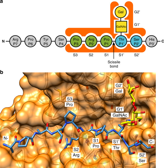
Akkermansia muciniphila is a mucin-degrading bacterium commonly found in the human gut that promotes a beneficial effect on health, likely based on the regulation of mucus thickness and gut barrier integrity, but also on the modulation of the immune system. In this work, we focus in OgpA from A. muciniphila, an O-glycopeptidase that exclusively hydrolyzes the peptide bond N-terminal to serine or threonine residues substituted with an O-glycan. We determine the high-resolution X-ray crystal structures of the unliganded form of OgpA, the complex with the glycodrosocin O-glycopeptide substrate and its product, providing a comprehensive set of snapshots of the enzyme along the catalytic cycle. In combination with O-glycopeptide chemistry, enzyme kinetics, and computational methods we unveil the molecular mechanism of O-glycan recognition and specificity for OgpA. The data also contribute to understanding how A. muciniphila processes mucins in the gut, as well as analysis of post-translational O-glycosylation events in proteins.
Conflict of interest statement
J.S. and A.N. are employees of Genovis A.B., and A.N. hold shares in the company. B.T., I.A., and M.E.G. declare no competing interests.
Figures


Similar articles
-
Structure-guided mutagenesis of a mucin-selective metalloprotease from Akkermansia muciniphila alters substrate preferences.J Biol Chem. 2022 May;298(5):101917. doi: 10.1016/j.jbc.2022.101917. Epub 2022 Apr 9. J Biol Chem. 2022. PMID: 35405095 Free PMC article.
-
A previously uncharacterized O-glycopeptidase from Akkermansia muciniphila requires the Tn-antigen for cleavage of the peptide bond.J Biol Chem. 2022 Oct;298(10):102439. doi: 10.1016/j.jbc.2022.102439. Epub 2022 Aug 30. J Biol Chem. 2022. PMID: 36049519 Free PMC article.
-
Unravelling the Glycan Code: Molecular Dynamics and Quantum Chemistry Reveal How O-Glycan Functional Groups Govern OgpA Selectivity in Mucin Degradation by Akkermansia muciniphila.Microb Biotechnol. 2025 Apr;18(4):e70091. doi: 10.1111/1751-7915.70091. Microb Biotechnol. 2025. PMID: 40181232 Free PMC article.
-
Akkermansia muciniphila plays critical roles in host health.Crit Rev Microbiol. 2023 Feb;49(1):82-100. doi: 10.1080/1040841X.2022.2037506. Epub 2022 May 21. Crit Rev Microbiol. 2023. PMID: 35603929 Review.
-
Disease-associated dysbiosis and potential therapeutic role of Akkermansia muciniphila, a mucus degrading bacteria of gut microbiome.Folia Microbiol (Praha). 2022 Dec;67(6):811-824. doi: 10.1007/s12223-022-00973-6. Epub 2022 May 20. Folia Microbiol (Praha). 2022. PMID: 35596115 Free PMC article. Review.
Cited by
-
The gut mucin-microbiota interactions: a missing key to optimizing endurance performance.Front Physiol. 2023 Nov 22;14:1284423. doi: 10.3389/fphys.2023.1284423. eCollection 2023. Front Physiol. 2023. PMID: 38074323 Free PMC article. Review.
-
A Bacteroides thetaiotaomicron genetic locus encodes activities consistent with mucin O-glycoprotein processing and N-acetylgalactosamine metabolism.Nat Commun. 2025 Apr 12;16(1):3485. doi: 10.1038/s41467-025-58660-2. Nat Commun. 2025. PMID: 40216766 Free PMC article.
-
Metaproteomics reveals parallel utilization of colonic mucin glycans and dietary fibers by the human gut microbiota.iScience. 2024 May 23;27(6):110093. doi: 10.1016/j.isci.2024.110093. eCollection 2024 Jun 21. iScience. 2024. PMID: 38947523 Free PMC article.
-
Sex hormones, intestinal inflammation, and the gut microbiome: Major influencers of the sexual dimorphisms in obesity.Front Immunol. 2022 Sep 27;13:971048. doi: 10.3389/fimmu.2022.971048. eCollection 2022. Front Immunol. 2022. PMID: 36248832 Free PMC article. Review.
-
Structure-guided mutagenesis of a mucin-selective metalloprotease from Akkermansia muciniphila alters substrate preferences.J Biol Chem. 2022 May;298(5):101917. doi: 10.1016/j.jbc.2022.101917. Epub 2022 Apr 9. J Biol Chem. 2022. PMID: 35405095 Free PMC article.
References
Publication types
MeSH terms
Substances
Supplementary concepts
Grants and funding
LinkOut - more resources
Full Text Sources
Molecular Biology Databases
