. 2020 Sep 24;10(1):15709.
doi: 10.1038/s41598-020-72748-3.

Topical ripasudil stimulates neuroprotection and axon regeneration in adult mice following optic nerve injury

Affiliations

Topical ripasudil stimulates neuroprotection and axon regeneration in adult mice following optic nerve injury

Euido Nishijima et al. Sci Rep. .

Abstract

Optic nerve injury induces optic nerve degeneration and retinal ganglion cell (RGC) death that lead to visual disturbance. In this study, we examined if topical ripasudil has therapeutic potential in adult mice after optic nerve crush (ONC). Topical ripasudil suppressed ONC-induced phosphorylation of p38 mitogen-activated protein kinase and ameliorated RGC death. In addition, topical ripasudil significantly suppressed the phosphorylation of collapsin response mediator protein 2 and cofilin, and promoted optic nerve regeneration. These results suggest that topical ripasudil promotes RGC protection and optic nerve regeneration by modulating multiple signaling pathways associated with neural cell death, microtubule assembly and actin polymerization.


Conflict of interest statement

The authors declare no competing interests.

Figures

Figure 1
Figure 1
Effects of topical ripasudil on RGC degeneration following ONC. (a) Experimental protocol. Ripasudil (2%; 5 µl) or PBS (5 µl) was administered locally every day, and the mice were euthanized at day 7 or 14 after ONC. (b) Schematic illustration of the retinal areas. (c) Immunostaining of the RBPMS-positive RGCs in the central, middle, and peripheral areas of the retina at 7 days after ONC. Scale bar: 50 µm. (df) Quantification of RGCs in the central (d), middle (e), and peripheral (f) areas of the retina. (g) Immunostaining of the RBPMS-positive RGCs in the central, middle, and peripheral areas of the retina at 14 days after ONC. Scale bar: 50 µm. (hj) Quantification of RGCs in the central (h), middle (i), and peripheral (j) areas of the retina. n = 4 per group. The data are presented as means ± SEM. *P < 0.05, **P < 0.01.
Figure 2
Figure 2
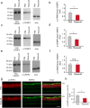
Effects of topical ripasudil on phosphorylation of p38 in the retinas following ONC. (a) Experimental protocol. Ripasudil (2%; 5 µl) or PBS (5 µl) was administered locally 3 min after ONC, and the mice were euthanized at 3 h and 1 day after ONC. (b) Immunoblot analysis of total p38, phosphorylated p38 (p-p38) and actin in the retinas at 0, 3 h and 1 day after ONC. Full length blot images are presented in Supplementary Fig. 1a. (c) Immunoblot analysis of total p38, p-p38 and actin in the retinas at 3 h after ONC. Full length blot images are presented in Supplementary Fig. 1b. (d) Quantitative analyses of (c). Ratio of p-p38 to total p38 in PBS-treated mice was estimated as 1.0. (e) Double-labeling immunohistochemistry of retinas with anti-p-p38 and anti-RBPMS antibodies. Scale bar: 50 µm. (f) Quantitative analyses of (e). The p-p38 intensity at the GCL in PBS-treated mice was estimated as 1.0. n = 4 per group. The data are presented as means ± SEM. *P < 0.05.
Figure 3
Figure 3
Effects of topical ripasudil on optic nerve regeneration following ONC. (a) Experimental protocol. Ripasudil (2%; 5 µl) or PBS (5 µl) was administered locally every day. CTB was intravitreally injected at day 12 and the mice were euthanized at day 14 after ONC. (b) Longitudinal sections through the optic nerve showing CTB-positive axons distal to the injury site (*) in PBS- and ripasudil-treated mice. Scale bar: 200 µm. (ce) Quantitative analyses of regenerating axons extending 100 (c), 250 (d), and 500 (e) µm from the injury site. n = 4 per group. The data are presented as means ± SEM. **P < 0.01, ***P < 0.001.
Figure 4
Figure 4
Expression of phosphorylated CRMP2 and cofilin in the retinas following ONC. (a) Immunoblot analysis of phosphorylated CRMP2 (p-CRMP2), total CRMP2, phosphorylated cofilin (p-cofilin), total cofilin and actin in the retina at 3 h, 1, 3, 7, and 14 days after ONC. Full length blot images are presented in Supplementary Fig. 1c and d. (b) Quantitative analyses of the ratio of p-CRMP2 to total CRMP2 in (a). Ratio of p-CRMP2 to total CRMP2 in control mice was estimated as 1.0. n = 9 (control), 3 (3 h, 1 and 3 days) or 10 (7 and 14 days). (c) Quantitative analyses of the ratio of p-cofilin to total cofilin in (a). Ratio of p-cofilin to total cofilin in control mice was estimated as 1.0. n = 3 per group. The data are presented as means ± SEM. *P < 0.05.
Figure 5
Figure 5
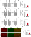
Effects of topical ripasudil on CRMP2 phosphorylation in the retinas after ONC. (a) Immunoblot analysis of total CRMP2 and p-CRMP2 in the retinas at 3 h after ONC. Full length blot images are presented in Supplementary Fig. 2a. (b) Quantitative analyses of (a). (c) Immunoblot analysis of total CRMP2 and p-CRMP2 in the retinas at 3 days after ONC. Full length blot images are presented in Supplementary Fig. 2b. (d) Quantitative analyses of (c). (e) Immunoblot analysis of total CRMP2 and p-CRMP2 in the retinas at 7 days after ONC. Full length blot images are presented in Supplementary Fig. 2c. (f) Quantitative analyses of (e). Ratio of p-CRMP2 to total CRMP2 in PBS-treated mice was estimated as 1.0. (g) Double-labeling immunohistochemistry of retinas with anti-p-CRMP2 and anti-RBPMS antibodies. Scale bar: 50 µm. (h) Quantitative analyses of (g). The p-CRMP2 intensity at the GCL in PBS-treated mice was estimated as 1.0. n = 4 per group. The data are presented as means ± SEM. *P < 0.05.
Figure 6
Figure 6
Effects of topical ripasudil on cofilin phosphorylation in the retinas after ONC. (a) Immunoblot analysis of total cofilin and p-cofilin in the retinas at 3 h after ONC. Full length blot images are presented in Supplementary Fig. 2d. (b) Quantitative analyses of (a). (c) Immunoblot analysis of total cofilin and p-cofilin in the retinas at 3 days after ONC. Full length blot images are presented in Supplementary Fig. 2e. (d) Quantitative analyses of (c). (e) Immunoblot analysis of total cofilin and p-cofilin in the retinas at 7 days after ONC. Full length blot images are presented in Supplementary Fig. 2f. (f) Quantitative analyses of (e). Ratio of p-cofilin to total cofilin in PBS-treated mice was estimated as 1.0. (g) Double-labeling immunohistochemistry of retinas with anti-p-cofilin and anti-RBPMS antibodies. Scale bar: 50 µm. (h) Quantitative analyses of (g). The p-cofilin intensity at the GCL in PBS-treated mice was estimated as 1.0. n = 4 per group. The data are presented as means ± SEM. *P < 0.05.

Similar articles

Cited by

References

    1. Katome T, et al. Inhibition of ASK1-p38 pathway prevents neural cell death following optic nerve injury. Cell Death Differ. 2013;20:270–280. doi: 10.1038/cdd.2012.122. - DOI - PMC - PubMed
    1. Noro T, et al. Spermidine promotes retinal ganglion cell survival and optic nerve regeneration in adult mice following optic nerve injury. Cell Death Dis. 2015;6:e1720. doi: 10.1038/cddis.2015.93. - DOI - PMC - PubMed
    1. Benowitz LI, He Z, Goldberg JL. Reaching the brain: advances in optic nerve regeneration. Exp. Neurol. 2017;287:365–373. doi: 10.1016/j.expneurol.2015.12.015. - DOI - PubMed
    1. Fujita Y, Yamashita T. Axon growth inhibition by RhoA/ROCK in the central nervous system. Front. Neurosci. 2014;8:338. doi: 10.3389/fnins.2014.00338. - DOI - PMC - PubMed
    1. Fournier AE, Takizawa BT, Strittmatter SM. Rho kinase inhibition enhances axonal regeneration in the injured CNS. J. Neurosci. 2003;23:1416–1423. doi: 10.1523/JNEUROSCI.23-04-01416.2003. - DOI - PMC - PubMed

Publication types

MeSH terms