

XBP1 negatively regulates CENPF expression via recruiting ATF6α to the promoter during ER stress
- PMID: 32973403
- PMCID: PMC7507253
- DOI: 10.1186/s12935-020-01553-9
XBP1 negatively regulates CENPF expression via recruiting ATF6α to the promoter during ER stress
Abstract
Background: Centromere protein F (CENPF) is a key component of the kinetochore complex involved in mitosis, cell differentiation and cellular response to stresses. However, the alteration of CENPF in response to endoplasmic reticulum (ER) stress has not been well described. In the present study, we investigate CENPF regulation in response to ER stress.
Methods: Quantitative real-time polymerase chain reaction and western blotting were used to determine CENPF expression under ER stress. Luciferase activity analysis was performed to investigate the promoter regions contributing to CENPF transcription in response to TG. Chromatin immunoprecipitation (ChIP) and ChIP Re-IP assays were used to determine if X-box binding protein 1 (XBP1) and/or activating transcription factor 6α (ATF6α) bind in the CENPF promoter region. Cell apoptosis and proliferation were analyzed using TUNEL, cell growth and clonogenic assays.
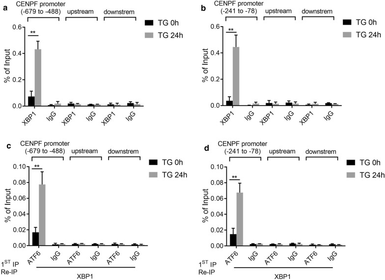
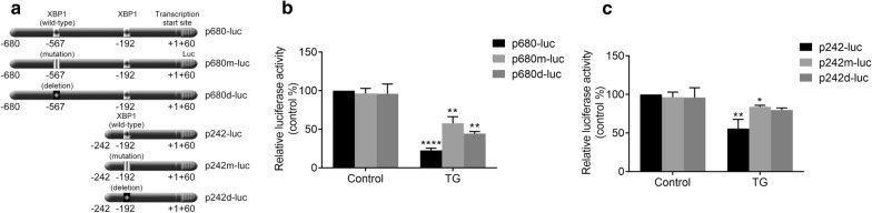
Results: CENPF expression is dramatically reduced under ER stress induced by thapsigargin (TG), brefeldin A (BFA), or tunicamycin (TM) and this downregulation of CENPF expression was dependent on XBP1 and ATF6α. Luciferase activity analysis of the truncated CENPF promoter indicates that regions from bases - 679 to - 488 and from - 241 to - 78 in the CENPF promoter were sensitive to TG treatment. Additionally, ChIP and ChIP Re-IP assays reveal that XBP1 and ATF6α were assembled on the same regions of CENPF promoter. Notably, we identify two XBP1 binding sequences at positions - 567 and - 192, to which XBP1 binding was enhanced by TG. Finally, CENPF overexpression inhibits cell apoptosis and promotes cell proliferation in response to ER stress.
Conclusion: In summary, these results demonstrate that ER stress plays a crucial role in CENPF expression, and XBP1 may up-regulate DNA-binding affinities after TG treatment to the promoter of CENPF. These findings may contribute to the understanding of the molecular mechanism of CENPF regulation.
Keywords: ATF6α; CENPF; ER stress; Expression regulation; XBP1.
© The Author(s) 2020.
Conflict of interest statement
Competing interestsThe authors declare that they have no competing interests.
Figures


Similar articles
-
Activating transcription factor 6 (ATF6) negatively regulates Polo-like kinase 4 expression via recruiting C/EBPβ to the upstream-promoter during ER stress.Biochim Biophys Acta Gene Regul Mech. 2020 Feb;1863(2):194488. doi: 10.1016/j.bbagrm.2020.194488. Epub 2020 Jan 9. Biochim Biophys Acta Gene Regul Mech. 2020. PMID: 31926341
-
CHOP negatively regulates Polo-like kinase 2 expression via recruiting C/EBPα to the upstream-promoter in human osteosarcoma cell line during ER stress.Int J Biochem Cell Biol. 2017 Aug;89:207-215. doi: 10.1016/j.biocel.2017.06.012. Epub 2017 Jun 24. Int J Biochem Cell Biol. 2017. PMID: 28652211
-
Identification of a novel endoplasmic reticulum stress response element regulated by XBP1.J Biol Chem. 2013 Jul 12;288(28):20378-91. doi: 10.1074/jbc.M113.457242. Epub 2013 Jun 4. J Biol Chem. 2013. PMID: 23737521 Free PMC article.
-
Endoplasmic reticulum stress induces PRNP prion protein gene expression in breast cancer.Breast Cancer Res. 2013 Mar 12;15(2):R22. doi: 10.1186/bcr3398. Breast Cancer Res. 2013. PMID: 23497519 Free PMC article.
-
Sledgehammer to Scalpel: Broad Challenges to the Heart and Other Tissues Yield Specific Cellular Responses via Transcriptional Regulation of the ER-Stress Master Regulator ATF6α.Int J Mol Sci. 2020 Feb 8;21(3):1134. doi: 10.3390/ijms21031134. Int J Mol Sci. 2020. PMID: 32046286 Free PMC article. Review.
Cited by
-
Centromere Protein F in Tumor Biology: Cancer's Achilles Heel.Cancer Med. 2025 May;14(10):e70949. doi: 10.1002/cam4.70949. Cancer Med. 2025. PMID: 40387105 Free PMC article. Review.
-
Identification of brain endothelial cell-specific genes and pathways in ischemic stroke by integrated bioinformatical analysis.Brain Circ. 2023 Nov 30;9(4):228-239. doi: 10.4103/bc.bc_40_23. eCollection 2023 Oct-Dec. Brain Circ. 2023. PMID: 38284111 Free PMC article.
-
Vernonia amygdalina Leaf Extract Induces Apoptosis in HeLa Cells: A Metabolomics and Proteomics Study.Pharmaceuticals (Basel). 2024 Aug 16;17(8):1079. doi: 10.3390/ph17081079. Pharmaceuticals (Basel). 2024. PMID: 39204184 Free PMC article.
-
Pivotal role of the endoplasmic reticulum stress-related XBP1s/miR-22/SIRT1 axis in acute myeloid leukemia apoptosis and response to chemotherapy.Leukemia. 2024 Aug;38(8):1764-1776. doi: 10.1038/s41375-024-02321-8. Epub 2024 Jun 22. Leukemia. 2024. PMID: 38909090 Free PMC article.
References
-
- Dai Y, Liu L, Zeng T, Zhu YH, Li J, Chen L, Li Y, Yuan YF, Ma S, Guan XY. Characterization of the oncogenic function of centromere protein F in hepatocellular carcinoma. Biochem Biophys Res Commun. 2013;436(4):711–718. - PubMed
LinkOut - more resources
Full Text Sources
Research Materials
Miscellaneous
