

NCS 613, a Potent PDE4 Inhibitor, Displays Anti-Inflammatory and Anti-Proliferative Properties on A549 Lung Epithelial Cells and Human Lung Adenocarcinoma Explants
- PMID: 32973507
- PMCID: PMC7466439
- DOI: 10.3389/fphar.2020.01266
NCS 613, a Potent PDE4 Inhibitor, Displays Anti-Inflammatory and Anti-Proliferative Properties on A549 Lung Epithelial Cells and Human Lung Adenocarcinoma Explants
Abstract
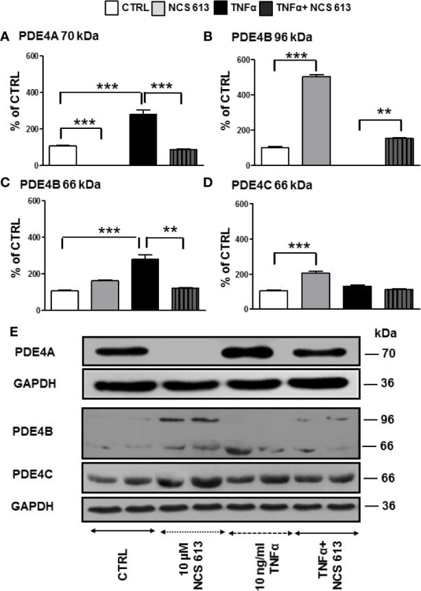
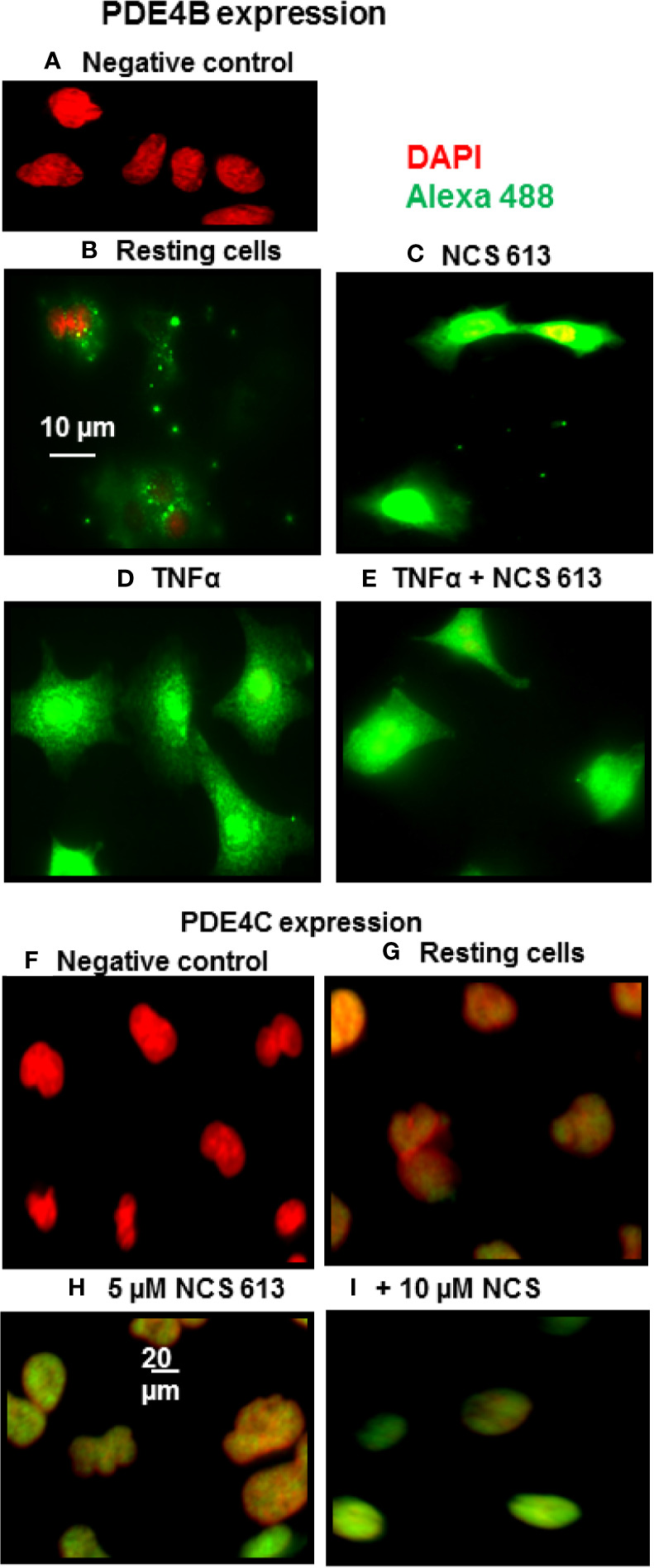
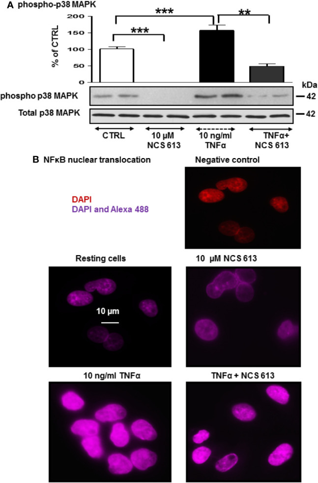
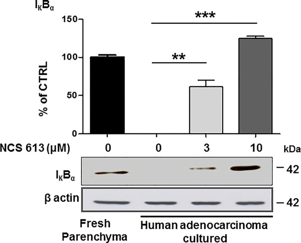
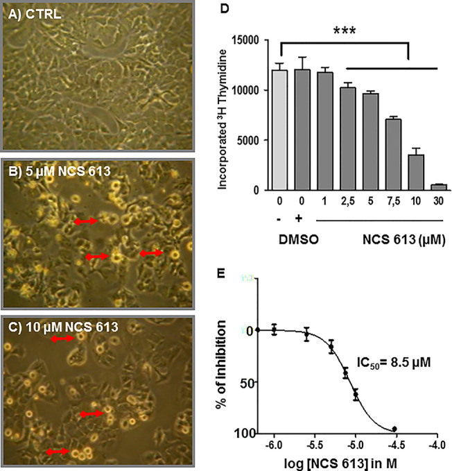
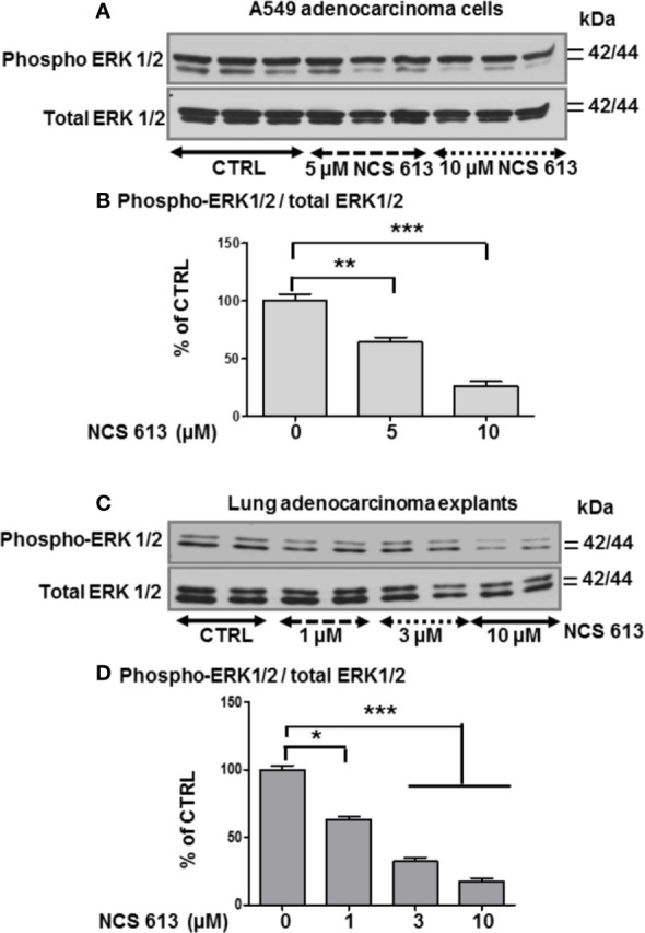
Chronic inflammation is a deleterious process occurring in several pulmonary diseases; it is a driving force promoting tumorigenesis. By regulating local cyclic nucleotide concentration, cyclic nucleotide phosphodiesterases (PDE) govern important biological processes, including inflammation and proliferation. The aim of this study was to investigate the anti-inflammatory and anti-proliferative effects of NCS 613, a specific PDE4 inhibitor, on TNFα-treated human lung adenocarcinoma cell line (A549) and on human lung adenocarcinoma explants. PDE4 isoforms and inflammatory pathways mediated by p38 MAPK, ERK1/2, and IκBα were analyzed by Western blot and immunostainings. Proliferation were performed using [3H]-thymidine incorporation under different experimental conditions. TNFα-stimulation increased p38 MAPK phosphorylation and NF-κB translocation into the nucleus, which was abolished by NCS 613 treatment. Concomitantly, NCS 613 restores IκBα detection level in human adenocarcinoma. An IC50 value of 8.5 μM was determined for NCS 613 on anti-proliferative properties while ERK1/2 signaling was down-regulated in A549 cells and lung adenocarcinoma explants. These findings shed light on PDE4 signaling as a key regulator of chronic inflammation and cancer epithelial cell proliferation. It suggests that PDE4 inhibition by NCS 613 represent potential and interesting strategy for therapeutic intervention in tackling chronic inflammation and cell proliferation.
Keywords: PDE4 inhibitor; cAMP signaling; human lung adenocarcinoma; inflammation; proliferation.
Copyright © 2020 Yougbare, Belemnaba, Morin, Abusnina, Senouvo, Keravis, Lugnier and Rousseau.
Figures
Similar articles
-
NCS 613, a potent and specific PDE4 inhibitor, displays anti-inflammatory effects on human lung tissues.Am J Physiol Lung Cell Mol Physiol. 2011 Oct;301(4):L441-50. doi: 10.1152/ajplung.00407.2010. Epub 2011 Jul 22. Am J Physiol Lung Cell Mol Physiol. 2011. PMID: 21784969
-
NCS 613 exhibits anti-inflammatory effects on PBMCs from lupus patients by inhibiting p38 MAPK and NF-κB signalling pathways while reducing proinflammatory cytokine production.Can J Physiol Pharmacol. 2013 May;91(5):353-61. doi: 10.1139/cjpp-2012-0233. Epub 2013 Jan 2. Can J Physiol Pharmacol. 2013. PMID: 23656347
-
Phosphodiesterase-4 promotes proliferation and angiogenesis of lung cancer by crosstalk with HIF.Oncogene. 2013 Feb 28;32(9):1121-34. doi: 10.1038/onc.2012.136. Epub 2012 Apr 23. Oncogene. 2013. PMID: 22525277
-
Phosphodiesterase inhibitors in airways disease.Eur J Pharmacol. 2006 Mar 8;533(1-3):110-7. doi: 10.1016/j.ejphar.2005.12.059. Epub 2006 Feb 2. Eur J Pharmacol. 2006. PMID: 16458289 Review.
-
PDE4 cAMP phosphodiesterases: modular enzymes that orchestrate signalling cross-talk, desensitization and compartmentalization.Biochem J. 2003 Feb 15;370(Pt 1):1-18. doi: 10.1042/BJ20021698. Biochem J. 2003. PMID: 12444918 Free PMC article. Review.
Cited by
-
Effects of pentoxifylline on mouse oocytes maturation and quality in vitro.Iran J Basic Med Sci. 2025;28(3):310-315. doi: 10.22038/ijbms.2024.77926.16856. Iran J Basic Med Sci. 2025. PMID: 39906611 Free PMC article.
-
Cyclic adenosine monophosphate/phosphodiesterase 4 pathway associated with immune infiltration and PD-L1 expression in lung adenocarcinoma cells.Front Oncol. 2022 Aug 1;12:904969. doi: 10.3389/fonc.2022.904969. eCollection 2022. Front Oncol. 2022. PMID: 35978822 Free PMC article.
-
Repurposing approved drugs for cancer therapy.Br Med Bull. 2021 Mar 25;137(1):13-27. doi: 10.1093/bmb/ldaa045. Br Med Bull. 2021. PMID: 33517358 Free PMC article. Review.
-
PDE4 inhibition as a therapeutic strategy for improvement of pulmonary dysfunctions in Covid-19 and cigarette smoking.Biochem Pharmacol. 2021 Mar;185:114431. doi: 10.1016/j.bcp.2021.114431. Epub 2021 Jan 28. Biochem Pharmacol. 2021. PMID: 33515531 Free PMC article. Review.
-
The multifaceted role of phosphodiesterase 4 in tumor: from tumorigenesis to immunotherapy.Front Immunol. 2025 Mar 10;16:1528932. doi: 10.3389/fimmu.2025.1528932. eCollection 2025. Front Immunol. 2025. PMID: 40129976 Free PMC article. Review.
References
-
- Abusnina A., Alhosin M., Keravis T., Muller C. D., Fuhrmann G., Bronner C., et al. (2011. a). Down-regulation of cyclic nucleotide phosphodiesterase PDE1A is the key event of p73 and UHRF1 deregulation in thymoquinone-induced acute lymphoblastic leukemia cell apoptosis. Cell Signal 23 (1), 152–160. 10.1016/j.cellsig.2010.08.015 - DOI - PubMed
-
- Alhosin M., Sharif T., Mousli M., Etienne-Selloum N., Fuhrmann G., Schini-Kerth V. B., et al. (2011). Down-regulation of UHRF1, associated with re-expression of tumor suppressor genes, is a common feature of natural compounds exhibiting anti-cancer pprperties. J. Exp. Clin. Cancer. Res. 30 (1), 41. 10.1186/1756-9966-30-41 - DOI - PMC - PubMed
LinkOut - more resources
Full Text Sources
Miscellaneous