Acute Hemorrhagic Leukoencephalitis: A Case and Systematic Review of the Literature
- PMID: 32973663
- PMCID: PMC7468463
- DOI: 10.3389/fneur.2020.00899
Acute Hemorrhagic Leukoencephalitis: A Case and Systematic Review of the Literature
Abstract
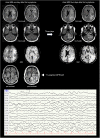
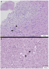
Objectives: To present a patient with acute hemorrhagic leukoencephalitis (AHLE) and a systematic review of the literature analyzing diagnostic procedures, treatment, and outcomes of AHLE. Methods: PubMed and Cochrane databases were screened. Papers published since 01/01/2000 describing adult patients are reported according to the PRISMA-guidelines. Results: A 59-year old male with rapidly developing coma and cerebral biopsy changes compatible with AHLE is presented followed by 43 case reports from the literature including males in 67% and a mean age of 38 years. Mortality was 47%. Infectious pathogens were reported in 35%, preexisting autoimmune diseases were identified in 12%. Neuroimaging revealed uni- or bihemispheric lesions in 65% and isolated lesions of the cerebellum, pons, medulla oblongata or the spinal cord without concomitant hemispheric involvement in 16%. Analysis of the cerebrospinal fluid showed an increased protein level in 87%, elevated white blood cells in 65%, and erythrocytes in 39%. Histology (reported in 58%) supported the diagnosis of AHLE in all cases. Glucocorticoids were used most commonly (97%), followed by plasmapheresis (26%), and intravenous immunoglobulins (12%), without a clear temporal relationship between treatment and the patients' clinical course. Conclusions: Although mortality was lower than previously reported, AHLE remains a life-threatening neurologic emergency with high mortality. Diagnosis is challenging as the level of evidence regarding the diagnostic yield of clinical, neuroimaging and laboratory characteristics remains low. Hence, clinicians are urged to heighten their awareness and to prompt cerebral biopsies in the context of rapidly progressive neurologic decline of unknown origin with the concurrence of the compiled characteristics. Future studies need to focus on treatment characteristics and their effects on course and outcome.
Keywords: immunosuppressive therapy; leukoencephalitis; mortality; outcome; parainfectious disease.
Copyright © 2020 Grzonka, Scholz, De Marchis, Tisljar, Rüegg, Marsch, Fladt and Sutter.
Figures


References
-
- Sureshbabu S, Babu R, Garg A, Peter S, Sobhana C, Mittal GK. Acute hemorrhagic leukoencephalitis unresponsive to aggressive immunosuppression. Clin Exp Neuroimmunol. (2017) 2017:63–6. 10.1111/cen3.12361 - DOI
Publication types
LinkOut - more resources
Full Text Sources

