Moderate Exercise in Spontaneously Hypertensive Rats Is Unable to Activate the Expression of Genes Linked to Mitochondrial Dynamics and Biogenesis in Cardiomyocytes
- PMID: 32973679
- PMCID: PMC7466645
- DOI: 10.3389/fendo.2020.00546
Moderate Exercise in Spontaneously Hypertensive Rats Is Unable to Activate the Expression of Genes Linked to Mitochondrial Dynamics and Biogenesis in Cardiomyocytes
Abstract
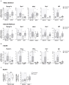
Hypertension (HTN) is a public health concern and a major preventable cause of cardiovascular disease (CVD). When uncontrolled, HTN may lead to adverse cardiac remodeling, left ventricular hypertrophy, and ultimately, heart failure. Regular aerobic exercise training exhibits blood pressure protective effects, improves myocardial function, and may reverse pathologic cardiac hypertrophy. These beneficial effects depend at least partially on improved mitochondrial function, decreased oxidative stress, endothelial dysfunction, and apoptotic cell death, which supports the general recommendation of moderate exercise in CVD patients. However, most of these mechanisms have been described on healthy individuals; the effect of moderate exercise on HTN subjects at a cellular level remain largely unknown. We hypothesized that hypertension in adult spontaneously hypertensive rats (SHRs) reduces the mitochondrial response to moderate exercise in the myocardium. Methods: Eight-month-old SHRs and their normotensive control-Wistar-Kyoto rats (WKYR)-were randomly assigned to moderate exercise on a treadmill five times per week with a running speed set at 10 m/min and 15° inclination. The duration of each session was 45 min with a relative intensity of 70-85% of the maximum O2 consumption for a total of 8 weeks. A control group of untrained animals was maintained in their cages with short sessions of 10 min at 10 m/min two times per week to maintain them accustomed to the treadmill. After completing the exercise protocol, we assessed maximum exercise capacity and echocardiographic parameters. Animals were euthanized, and heart and muscle tissue were harvested for protein determinations and gene expression analysis. Measurements were compared using a nonparametric ANOVA (Kruskal-Wallis), with post-hoc Dunn's test. Results: At baseline, SHR presented myocardial remodeling evidenced by left ventricular hypertrophy (interventricular septum 2.08 ± 0.07 vs. 1.62 ± 0.08 mm, p < 0.001), enlarged left atria (0.62 ± 0.1 mm vs. 0.52 ± 0.1, p = 0.04), and impaired diastolic function (E/A ratio 2.43 ± 0.1 vs. 1.56 ± 0.2) when compared to WKYR. Moderate exercise did not induce changes in ventricular remodeling but improved diastolic filling pattern (E/A ratio 2.43 ± 0.1 in untrained SHR vs. 1.89 ± 0.16 trained SHR, p < 0.01). Histological analysis revealed increased myocyte transversal section area, increased Myh7 (myosin heavy chain 7) expression, and collagen fiber accumulation in SHR-control hearts. While the exercise protocol did not modify cardiac size, there was a significant reduction of cardiomyocyte size in the SHR-exercise group. Conversely, titin expression increased only WYK-exercise animals but remained unchanged in the SHR-exercise group. Mitochondrial response to exercise also diverged between SHR and WYKR: while moderate exercise showed an apparent increase in mRNA levels of Ppargc1α, Opa1, Mfn2, Mff, and Drp1 in WYKR, mitochondrial dynamics proteins remained unchanged in response to exercise in SHR. This finding was further confirmed by decreased levels of MFN2 and OPA1 in SHR at baseline and increased OPA1 processing in response to exercise in heart. In summary, aerobic exercise improves diastolic parameters in SHR but fails to activate the cardiomyocyte mitochondrial adaptive response observed in healthy individuals. This finding may explain the discrepancies on the effect of exercise in clinical settings and evidence of the need to further refine our understanding of the molecular response to physical activity in HTN subjects.
Keywords: cardiac remodeling; exercise; heart; hypertension; mitochondrial dynamics.
Copyright © 2020 Quiroga, Mancilla, Oyarzun, Tapia, Caballero, Gabrielli, Valladares-Ide, del Campo, Castro and Verdejo.
Figures


Similar articles
-
Left ventricular remodeling with exercise in hypertension.Am J Physiol Heart Circ Physiol. 2009 Oct;297(4):H1361-8. doi: 10.1152/ajpheart.01253.2008. Epub 2009 Aug 7. Am J Physiol Heart Circ Physiol. 2009. PMID: 19666835 Free PMC article.
-
High-intensity intermittent training is as effective as moderate continuous training, and not deleterious, in cardiomyocyte remodeling of hypertensive rats.J Appl Physiol (1985). 2019 Apr 1;126(4):903-915. doi: 10.1152/japplphysiol.00131.2018. Epub 2019 Jan 31. J Appl Physiol (1985). 2019. PMID: 30702976
-
Cardiac benefits of exercise training in aging spontaneously hypertensive rats.J Hypertens. 2011 Dec;29(12):2349-58. doi: 10.1097/HJH.0b013e32834d2532. J Hypertens. 2011. PMID: 22045123
-
The ageing spontaneously hypertensive rat as a model of the transition from stable compensated hypertrophy to heart failure.Eur Heart J. 1995 Dec;16 Suppl N:19-30. doi: 10.1093/eurheartj/16.suppl_n.19. Eur Heart J. 1995. PMID: 8682057 Review.
-
Cardiac changes in spontaneously hypertensive rats: Modulation by aerobic exercise.Prog Biophys Mol Biol. 2023 Jan;177:109-124. doi: 10.1016/j.pbiomolbio.2022.11.001. Epub 2022 Nov 5. Prog Biophys Mol Biol. 2023. PMID: 36347337 Review.
Cited by
-
Human myocardial mitochondrial oxidative capacity is impaired in mild acute heart transplant rejection.ESC Heart Fail. 2021 Dec;8(6):4674-4684. doi: 10.1002/ehf2.13607. Epub 2021 Sep 6. ESC Heart Fail. 2021. PMID: 34490749 Free PMC article.
-
Regulation of Mitochondrial Dynamics by Aerobic Exercise in Cardiovascular Diseases.Front Cardiovasc Med. 2022 Jan 13;8:788505. doi: 10.3389/fcvm.2021.788505. eCollection 2021. Front Cardiovasc Med. 2022. PMID: 35097008 Free PMC article. Review.
-
The structure and function of mitofusin 2 and its role in cardiovascular disease through mediating mitochondria-associated endoplasmic reticulum membranes.Front Cardiovasc Med. 2025 May 30;12:1535401. doi: 10.3389/fcvm.2025.1535401. eCollection 2025. Front Cardiovasc Med. 2025. PMID: 40520935 Free PMC article. Review.
-
Current Understanding of the Pivotal Role of Mitochondrial Dynamics in Cardiovascular Diseases and Senescence.Front Cardiovasc Med. 2022 May 18;9:905072. doi: 10.3389/fcvm.2022.905072. eCollection 2022. Front Cardiovasc Med. 2022. PMID: 35665261 Free PMC article. Review.
-
Time of day of exercise does not affect the beneficial effect of exercise on bone structure in older female rats.Front Physiol. 2023 Oct 26;14:1142057. doi: 10.3389/fphys.2023.1142057. eCollection 2023. Front Physiol. 2023. PMID: 37965104 Free PMC article.
References
Publication types
MeSH terms
LinkOut - more resources
Full Text Sources
Medical
Miscellaneous

