

Enterovirus Replication Organelles and Inhibitors of Their Formation
- PMID: 32973693
- PMCID: PMC7468505
- DOI: 10.3389/fmicb.2020.01817
Enterovirus Replication Organelles and Inhibitors of Their Formation
Abstract
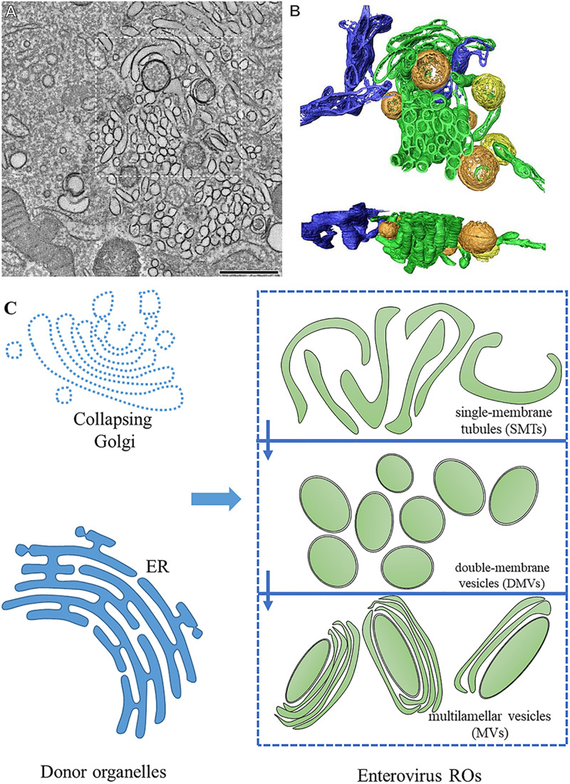
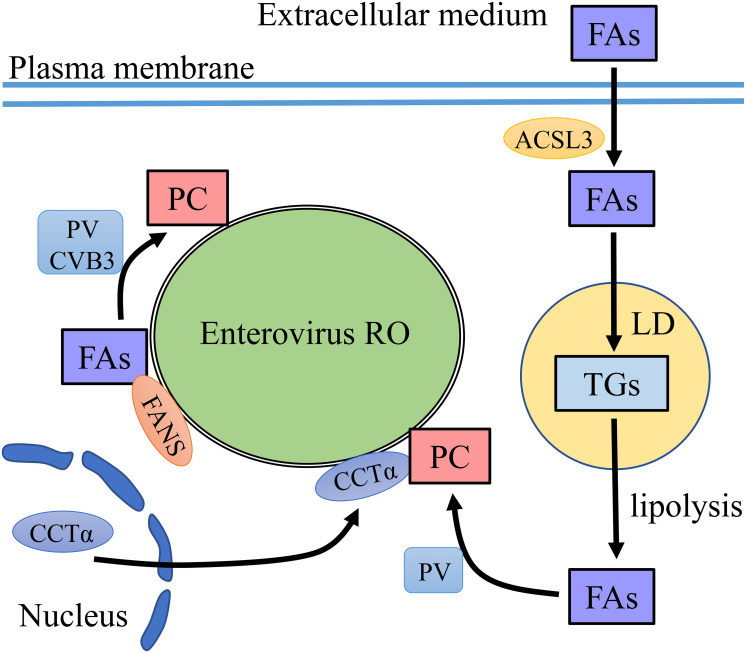
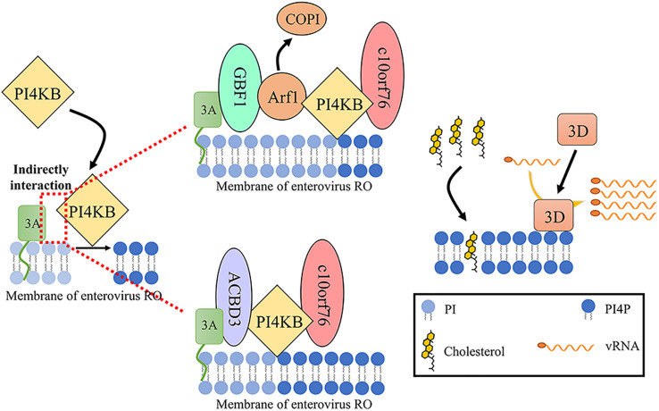
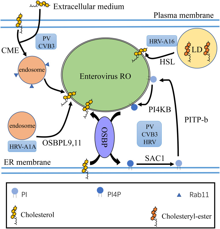
Enteroviral replication reorganizes the cellular membrane. Upon infection, viral proteins and hijacked host factors generate unique structures called replication organelles (ROs) to replicate their viral genomes. ROs promote efficient viral genome replication, coordinate the steps of the viral replication cycle, and protect viral RNA from host immune responses. More recent researches have focused on the ultrastructure structures, formation mechanism, and functions in the virus life cycle of ROs. Dynamic model of enterovirus ROs structure is proposed, and the secretory pathway, the autophagy pathway, and lipid metabolism are found to be associated in the formation of ROs. With deeper understanding of ROs, some compounds have been found to show inhibitory effects on viral replication by targeting key proteins in the process of ROs formation. Here, we review the recent findings concerning the role, morphology, biogenesis, formation mechanism, and inhibitors of enterovirus ROs.
Keywords: biogenesis; enteroviruses; inhibitors; lipid metabolism; replication organelles.
Copyright © 2020 Li, Wang, Cheng, Wen, Ou, Mao, Gao, Sun, Jia, Yang, Wu, Zhu, Zhao, Chen, Liu, Zhang, Liu, Yu, Zhang, Tian, Pan and Chen.
Figures
Similar articles
-
The Origin, Dynamic Morphology, and PI4P-Independent Formation of Encephalomyocarditis Virus Replication Organelles.mBio. 2018 Apr 17;9(2):e00420-18. doi: 10.1128/mBio.00420-18. mBio. 2018. PMID: 29666283 Free PMC article.
-
ACBD3 Is an Essential Pan-enterovirus Host Factor That Mediates the Interaction between Viral 3A Protein and Cellular Protein PI4KB.mBio. 2019 Feb 12;10(1):e02742-18. doi: 10.1128/mBio.02742-18. mBio. 2019. PMID: 30755512 Free PMC article.
-
Escaping Host Factor PI4KB Inhibition: Enterovirus Genomic RNA Replication in the Absence of Replication Organelles.Cell Rep. 2017 Oct 17;21(3):587-599. doi: 10.1016/j.celrep.2017.09.068. Cell Rep. 2017. PMID: 29045829 Free PMC article.
-
Fat(al) attraction: Picornaviruses Usurp Lipid Transfer at Membrane Contact Sites to Create Replication Organelles.Trends Microbiol. 2016 Jul;24(7):535-546. doi: 10.1016/j.tim.2016.02.017. Epub 2016 Mar 22. Trends Microbiol. 2016. PMID: 27020598 Free PMC article. Review.
-
The interplay of autophagy and enterovirus.Semin Cell Dev Biol. 2020 May;101:12-19. doi: 10.1016/j.semcdb.2019.08.001. Epub 2019 Sep 25. Semin Cell Dev Biol. 2020. PMID: 31563390 Free PMC article. Review.
Cited by
-
Viral Conjunctivitis.Viruses. 2023 Mar 4;15(3):676. doi: 10.3390/v15030676. Viruses. 2023. PMID: 36992385 Free PMC article. Review.
-
Coxsackievirus Protease 2A Targets Host Protease ATG4A to Impair Autophagy.Viruses. 2022 Sep 13;14(9):2026. doi: 10.3390/v14092026. Viruses. 2022. PMID: 36146840 Free PMC article.
-
Repurposing Anticancer Drugs Targeting the MAPK/ERK Signaling Pathway for the Treatment of Respiratory Virus Infections.Int J Mol Sci. 2024 Jun 25;25(13):6946. doi: 10.3390/ijms25136946. Int J Mol Sci. 2024. PMID: 39000055 Free PMC article. Review.
-
Direct-Acting Antivirals and Host-Targeting Approaches against Enterovirus B Infections: Recent Advances.Pharmaceuticals (Basel). 2023 Jan 29;16(2):203. doi: 10.3390/ph16020203. Pharmaceuticals (Basel). 2023. PMID: 37259352 Free PMC article. Review.
-
Non-lytic spread of poliovirus requires the nonstructural protein 3CD.mBio. 2025 Jan 8;16(1):e0327624. doi: 10.1128/mbio.03276-24. Epub 2024 Dec 12. mBio. 2025. PMID: 39665531 Free PMC article.
References
Publication types
LinkOut - more resources
Full Text Sources
Other Literature Sources