. 2020 Aug 20:11:1955.
doi: 10.3389/fmicb.2020.01955. eCollection 2020.

Phagolysosomal Survival Enables Non-lytic Hyphal Escape and Ramification Through Lung Epithelium During Aspergillus fumigatus Infection

Affiliations

Phagolysosomal Survival Enables Non-lytic Hyphal Escape and Ramification Through Lung Epithelium During Aspergillus fumigatus Infection

Constanze Seidel et al. Front Microbiol. .

Abstract

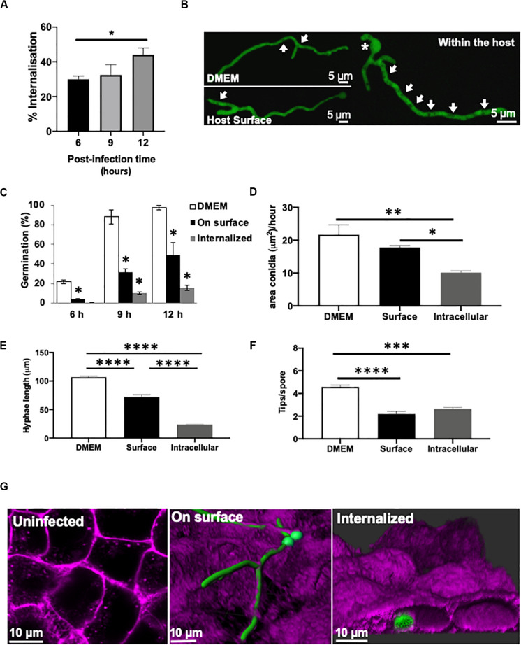
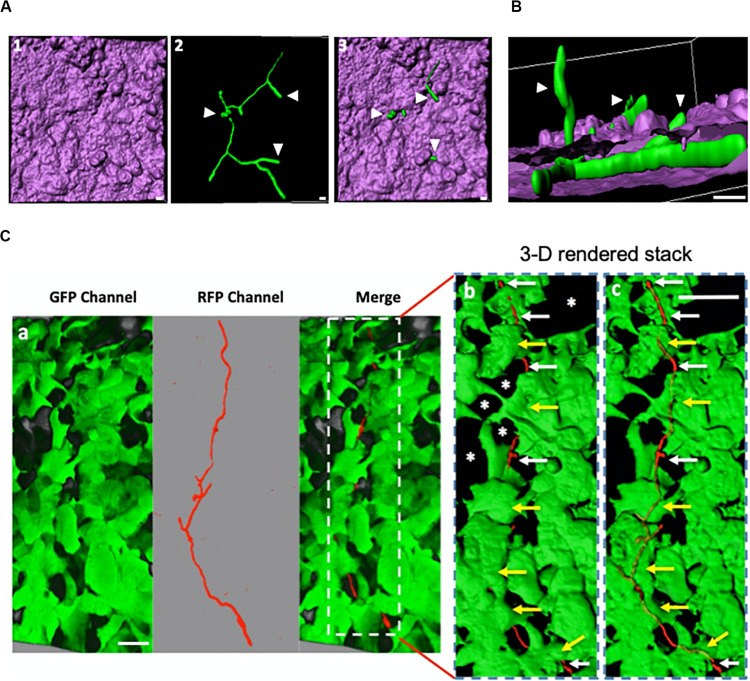
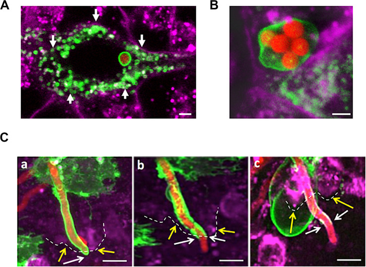
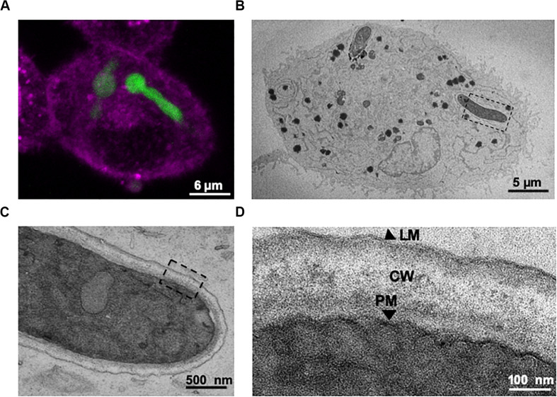
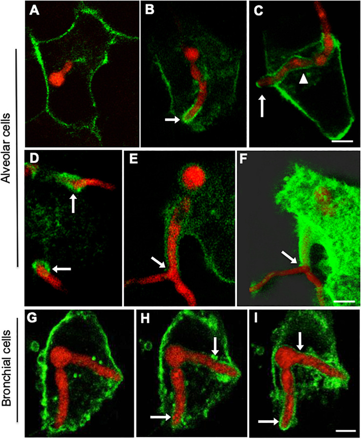
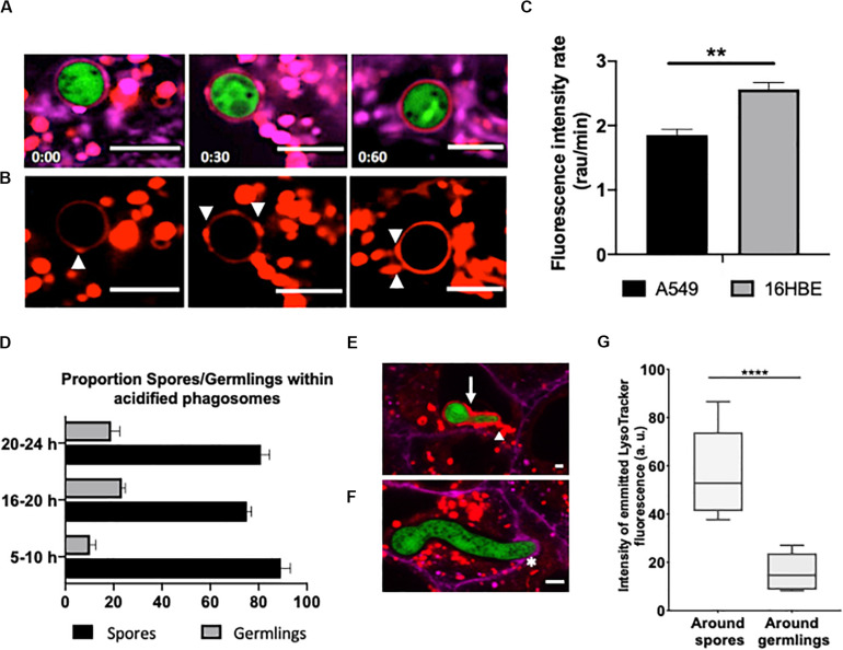
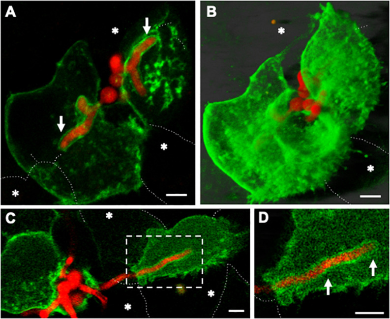
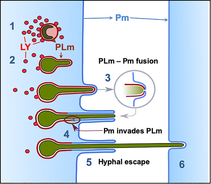
Aspergillus fumigatus is the most important mould pathogen in immunosuppressed patients. Suboptimal clearance of inhaled spores results in the colonisation of the lung airways by invasive hyphae. The first point of contact between A. fumigatus and the host is the lung epithelium. In vitro and ex vivo studies have characterised critical aspects of the interaction of invasive hyphae on the surface of epithelial cells. However, the cellular interplay between internalised A. fumigatus and the lung epithelium remains largely unexplored. Here, we use high-resolution live-cell confocal microscopy, 3D rendered imaging and transmission electron microscopy to define the development of A. fumigatus after lung epithelium internalisation in vitro. Germination, morphology and growth of A. fumigatus were significantly impaired upon internalisation by alveolar (A549) and bronchial (16HBE) lung epithelial cells compared to those growing on the host surface. Internalised spores and germlings were surrounded by the host phagolysosome membrane. Sixty per cent of the phagosomes containing germlings were not acidified at 24 h post infection allowing hyphal development. During escape, the phagolysosomal membrane was not ruptured but likely fused to host plasma membrane allowing hyphal exit from the intact host cell in an non-lytic Manner. Subsequently, escaping hyphae elongated between or through adjacent epithelial lung cells without penetration of the host cytoplasm. Hyphal tips penetrating new epithelial cells were surrounded by the recipient cell plasma membrane. Altogether, our results suggest cells of lung epithelium survive fungal penetration because the phagolysosomal and plasma membranes are never breached and that conversely, fungal spores survive due to phagosome maturation failure. Consequently, fungal hyphae can grow through the epithelial cell layer without directly damaging the host. These processes likely prevent the activation of downstream immune responses alongside limiting the access of professional phagocytes to the invading fungal hypha. Further research is needed to investigate if these events also occur during penetration of fungi in endothelial cells, fibroblasts and other cell types.

Keywords: Aspergillus fumigatus; airway epithelium; aspergillosis; epithelial cell infection; membrane compartmentalisation; microscopy; phagolysosome.


Figures

FIGURE 1
FIGURE 1
Germination and morphology of internalised A. fumigatus is impaired during infection of type II alveolar cells. (A) A. fumigatus spore uptake by A549 epithelial cells. (B) Live-cell imaging of hyphae of A. fumigatus expressing cytoplasmic GFP in DMEM, on the alveolar host surface or within the host cells at 37°C, 5% CO2 for 18 h in the absence of nystatin. Arrows indicate vacuoles. Asterisks highlight sites of second germ tube initiation. A. fumigatus (C) germination rate (%), (D) swelling rate (%), (E) hyphal extension rate and (F) degree of branching in the presence or absence of A549 alveolar cells (G) Live-cell imaging of the different strategies of A. fumigatus infections of A549 alveolar cells. Magenta = host’s plasma membrane stained with CellMask deep red. Green = A. fumigatus expressing cytosolic GFP. Measurements were performed in three biological and technical replicates (average ± standard deviation [SD]). P < 0.05; ∗∗P < 0.01; ∗∗∗P < 0.001; ****P < 0.0001.
FIGURE 2
FIGURE 2
Internalised A. fumigatus can expand between adjacent epithelial cells. (A) Live-cell imaging of hyphae of A. fumigatus cytoplasmically labelled with GFP infecting a monolayer of A549 alveolar cells cultured in DMEM at 37°C, 5% CO2 for 18 h. 1- 3-D rendered image of an infected alveolar monolayer stained with Cell Mask Deep Red. 2- Internalised A. fumigatus hyphae 3- Merge of 1 and 2. Scale bars: 5 μm. (B) Lateral view of the 3-D confocal stack displayed in (A) Arrowheads indicate hyphal tips escaping the host monolayer Scale bar: 5 μm. (C) A. fumigatus hyphal (red) invasion of the epithelial cells (green) 18 h post-inoculation, a magnification of the part indicated with dashed white rectangle with surface rendering, and with 60% transparency demonstrating the internal invading hypha. White arrows indicate hypha that have emerged or growth through unlabelled (asterisks) cells and yellow arrows indicate intracellular parts of the hypha. Scale bars: 40 μm.
FIGURE 3
FIGURE 3
A. fumigatus conidia are surrounded by a phagolysosomal membrane upon internalisation, germination and during A549 escape. (A) Single internalised conidia surrounded by a phagolysosomal membrane. A large number of individual lysosomes are accumulated in proximity to the host cell membrane (arrows). (B) Multiple internalised conidia surrounded by a single phagolysosomal compartment. (C) Time course of escaping of an extending hyphae from the phagolysosome demonstrates this process to involve (a) stretching of the phagosome; (b) escape from the host cell and (c) escape from the phagolysosome. Yellow arrows and dashes indicate the host cytoplasmic membrane; white arrows indicate the distal part of the tip escaping from the phagolysosome (Scale bars = 5 μm). Red = A. fumigatus expressing cytoplasmic RFP; Green = phagolysosomal compartment stained with LAMP-1 GFP. Magenta = A549 cell plasma membrane stained with CellMask deep red.
FIGURE 4
FIGURE 4
Correlative live-cell imaging and transmission electron microscopy (TEM) confirms that internalised A. fumigatus growing germlings are engulfed within the lung epithelium phagosome. (A) Confocal live-cell imaging of an internalised germling by A549 epithelial cells. (B) TEM image of a cross-section of the same infected A549 cell. (C) Magnification of the tip of the internalised germling. (D) Sections of the germling cell wall, PM: A. fumigatus plasma membrane; CW: A. fumigatus cell wall; LM: host phagolysosomal membrane. Note the host’s lipid bilayer that surrounds A. fumigatus within A549 host cells shown in panel (D).
FIGURE 5
FIGURE 5
Internalised A. fumigatus cells are surrounded by the host’s plasma membrane upon escape of alveolar and bronchial epithelial cells. Confocal live-cell imaging shows A. fumigatus expressing cytoplasmic RFP within A549 and 16HBE epithelial cells expressing GFP in the plasma membrane. (A) Internalised germling before reaching the host plasma membrane. (B) The tip (arrow) of the internalised germlings is surrounded by host plasma membrane during early interaction. (C) After late interaction of the internalised germling tip, the host plasma membrane surrounds the basal part of the germling (arrowhead). (D) Host plasma membrane is accumulated in the regions of hyphal escape (arrows). (E) Host plasma membrane surrounds an extension of the escaping germling. (F) Maximal projection shown in panel (E). (G–I) 16HBE plasma membrane (arrows) surrounds internalised A. fumigatus germlings upon escape. Scale bars = 5 μm.
FIGURE 6
FIGURE 6
Escaping A. fumigatus germlings are surrounded by a non-acidic phagosomal compartment. Alveolar monolayers were challenged with A. fumigatus (green) for 18 h at 37°C, 5% CO2. Following incubation times, cells were stained with Cell Mask Deep Red (magenta) and LysoTracker (red). (A) Time lapse (min) of an internalised spore surrounded by an acidic phagosome. (B) LysoTracker channel shown in A. Arrowheads highlight the site of lysosome fusion with the phagosome. (C) Acidification rate of spores internalised by either alveolar or bronchial cells. r.a.u estates for relative abundance units. (D) Time-lapse quantification of the percentage of germlings surrounded by acidified and non-acidified phagosomes. (E) Merged images of an internalised young germling. The arrow highlights the acidic compartment that surrounds A. fumigatus. The arrowhead highlights the fusion site of the acidic compartments. (F) Merged images of an internalised mature germling. The asterisk highlights the initial interaction between the mature germling tip and the protrusion of the host plasma membrane. Note there is no fusion of acidic compartments surrounding A. fumigatus. (G) Relative fluorescence of LysoTracker in internalised spores and germlings. Measurements were performed three times on three biological samples (average ± standard deviation [SD]) (**P < 0.01 and ****P < 0.0001). Scale bars: 5 μm.
FIGURE 7
FIGURE 7
Host plasma membrane compartmentalisation of A. fumigatus hyphae occurs upon invasion of naïve lung epithelial neighbouring cells. Confocal live-cell imaging of A. fumigatus expressing cytoplasmic RFP within a GFP-plasma membrane transfected A549 epithelial cell line at 37°C, 5% CO2. (A) Germ-tubes of internalised spores extend toward adjacent cells. (B) Maximal projection of the same cells shown in A. (C) Upon escape of an epithelial host the apical zone of an invading leading hyphae is surrounded by the plasma membrane of a naïve host cell. (D) Zoom in of the apical region of the hypha shown in (A). White arrows indicate the plasma membrane of the new host. Asterisks highlight epithelial cells that were not transfected. Scale bars: 5 μm.
FIGURE 8
FIGURE 8
Phagolysosome–Plasma membrane dynamics during A. fumigatus escape. (1) Most A. fumigatus conidia are killed by acidification of phagosomes and lysosomal fusion, (2) In some circumstances lysosome recruitment to the phagosome containing A. fumigatus conidia does not inhibit fungal growth and acidification does not occur allowing conidial germination. (3) The PLm continuously extends while surrounding the intracellular growing germling until the PLm contacts the plasma membrane. (4) The PLm and Pm fuse and (5) the host plasma membrane migrates into the phagolysosome membrane facilitating hyphal escape. (6) Hyphae then proceed to penetrate further host cells without rupture of the plasma membrane. LY: lysosome, PL: phagolysosome, PLm; phagolysosome membrane, Pm: plasma membrane.

Similar articles

Cited by

References

    1. Alpuche-Aranda C. M., Racoosin E. L., Swanson J. A., Miller S. I. (1994). Salmonella stimulate macrophage macropinocytosis and persist within spacious phagosomes. J. Exp. Med. 179 601–608. 10.1084/jem.179.2.601 - DOI - PMC - PubMed
    1. Amich J., Mokhtari Z., Strobel M., Vialetto E., Sheta D., Yu Y., et al. (2020). Three-dimensional light sheet fluorescence microscopy of lungs to dissect local host immune-Aspergillus fumigatus interactions. mBio 11:e02752-9. 10.1128/mBio.02752-19 - DOI - PMC - PubMed
    1. Amin S., Thywissen A., Heinekamp T., Saluz H. P., Brakhage A. A. (2014). Melanin dependent survival of Apergillus fumigatus conidia in lung epithelial cells. Int. J. Med. Microbiol. 304 626–636. 10.1016/j.ijmm.2014.04.009 - DOI - PubMed
    1. Araujo R., Rodrigues A. G. (2004). Variability of germinative potential among pathogenic species of Aspergillus. J. Clin. Microbiol. 42 4335–4337. 10.1128/JCM.42.9.4335-4337.2004 - DOI - PMC - PubMed
    1. Ballard E., Melchers W. J. G., Zoll J., Brown A. J. P., Verweij P. E., Warris A. (2018). In-host microevolution of Aspergillus fumigatus: a phenotypic and genotypic analysis. Fungal Genet. Biol. 113 1–13. 10.1016/j.fgb.2018.02.003 - DOI - PMC - PubMed

LinkOut - more resources