

Interstitial Lung Disease in Children With Selected Primary Immunodeficiency Disorders-A Multicenter Observational Study
- PMID: 32973798
- PMCID: PMC7481462
- DOI: 10.3389/fimmu.2020.01950
Interstitial Lung Disease in Children With Selected Primary Immunodeficiency Disorders-A Multicenter Observational Study
Abstract
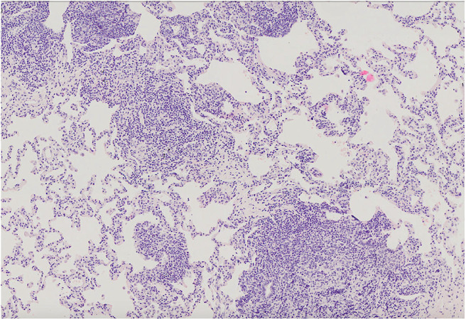
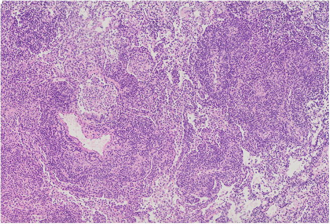
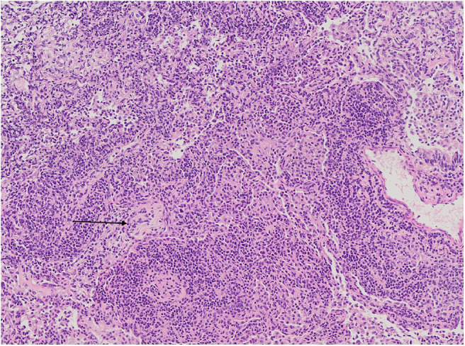
Primary immunodeficiencies (PIDs) are rare disorders of the immune system encompassing inborn errors of immunity. Primary antibody deficiencies constitute the largest group of PID with common variable immunodeficiency (CVID) being the most common symptomatic form. Combined immunodeficiencies (CID) accompanied by antibody deficiency can mimic CVID and these patients need the verification of the final diagnosis. Respiratory involvement, especially interstitial lung disease (ILD), poses a relevant cause of morbidity and mortality among patients with PID and in some cases is the first manifestation of immunodeficiency. In this study we present a retrospective analysis of a group of children with primary immunodeficiency and ILD - the clinical, radiological, histological characteristics, treatment strategies and outcomes. Eleven children with PID-related ILD were described. The majority of them presented CVID, in three patients CID was recognized. All patients underwent detailed pulmonary diagnostics. In eight of them histological analysis of lung biopsy was performed. We noted that in two out of 11 patients acute onset of ILD with respiratory failure was the first manifestation of the disease and preceded PID diagnosis. The most common histopathological diagnosis was GLILD. Among the analyzed patients three did not require any immunosuppressive therapy. All eight treated children received corticosteroids as initial treatment, but in some of them second-line therapy was introduced. The relevant side effects in some patients were observed. The study demonstrated that the response to corticosteroids is usually prompt. However, the resolution of pulmonary changes may be incomplete and second-line treatment may be necessary.
Keywords: CVID; GLILD; children; computed tomography; interstitial lung disease; primary immunodeficiency.
Copyright © 2020 Pac, Bielecka, Grzela, Komarnicka, Langfort, Koltan, Dabrowska-Leonik, Bernat-Sitarz, Pronicki, Dmenska, Pituch-Noworolska, Mikoluc, Piatosa, Tkaczyk, Bernatowska, Wojsyk-Banaszak and Krenke.
Figures
References
-
- Tangye SG, Al-Herz W, Bousfiha A, Chatila T, Cunningham-Rundles C, Etzioni A, et al. Human inborn errors of immunity: 2019 update on the classification from the International Union of Immunological Societies Expert Committee. J Clin Immunol. (2020) 40:66–81. 10.1007/s10875-019-00737-x - DOI - PMC - PubMed
Publication types
MeSH terms
Substances
LinkOut - more resources
Full Text Sources
Medical