Case Reports
. 2020 Aug 21:11:938.
doi: 10.3389/fgene.2020.00938. eCollection 2020.

Atypical Retinal Phenotype in a Patient With Alström Syndrome and Biallelic Novel Pathogenic Variants in ALMS1, Including a de novo Variation

Affiliations
Case Reports

Atypical Retinal Phenotype in a Patient With Alström Syndrome and Biallelic Novel Pathogenic Variants in ALMS1, Including a de novo Variation

Laura Mauring et al. Front Genet. .

Abstract

Alström syndrome (ALMS) is a rare autosomal recessive multi-organ syndrome considered to date as a ciliopathy and caused by variations in ALMS1. Phenotypic variability is well-documented, particularly for the systemic disease manifestations; however, early-onset progressive retinal degeneration affecting both cones and rods (cone-rod type) is universal, leading to blindness by the teenage years. Other features include cardiomyopathy, kidney dysfunction, sensorineural deafness, and childhood obesity associated with hyperinsulinemia and type 2 diabetes mellitus. Here, we present an unusual and delayed retinal dystrophy phenotype associated with ALMS in a 14-year-old female, with affected cone function and surprising complete preservation of rod function on serial electroretinograms (ERGs). High-throughput sequencing of the affected proband revealed compound heterozygosity with two novel nonsense variations in the ALMS1 gene, including one variant of de novo inheritance, an unusual finding in autosomal recessive diseases. To confirm the diagnosis in the context of an unusually mild phenotype and identification of novel variations, we demonstrated the biallelic status of the compound heterozygous variations (c.[286C > T];[1211C > G], p.[(Gln96*)];[(Ser404*)]). This unique case extends our knowledge of the phenotypic variability and the pathogenic variation spectrum in ALMS patients.

Keywords: ALMS1 gene; Alström syndrome; de novo variation; high throughput sequencing; retinal dystrophy.


Figures

Figure 1
Figure 1
Fundus photography, fundus autofluorescence, and optical coherence scanning images (OCT) of the patient’s retina. Fundus photography of right (A) and left (B) eyes demonstrates a granular appearance of the macula bilaterally (white arrows). No attenuation of retinal blood vessels can be observed. Fundus autofluorescence of right eye (C) and left eye (D) demonstrates increased autofluorescence of the fovea bilaterally (white arrows). OCT of right (E) and left (F) eye shows irregularity of the ellipsoid zone as well as an interdigitation zone in the macular region (white arrows).
Figure 2
Figure 2
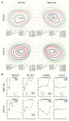
Kinetic visual fields and electroretinogram (ERG) results. (A) Kinetic visual fields of the patient’s eyes shows marked central loss of sensitivity with some constriction of the peripheral visual field compared to the reference (control). (B) Full field ERG of both eyes were performed according to the ISCEV (International Society for clinical Electrophysiology of vision) standard. Undetectable full field cone ERGs (light adapted LA 3.0 and LA 30 Hz) with subnormal dark adapted (DA 0.01 rod specific) and bright flash dark adapted (DA 10.0) compared to the reference (control is shown as small-integrated panels within each upper panels) is demonstrated.
Figure 3
Figure 3
Identification of two pathogenic variations in ALMS1. (A) Targeted exome sequencing reads aligned to exon 1 and 5 of the ALMS1 gene, displaying the two heterozygous variations in IGV (Thorvaldsdóttir et al., 2012). (B) Pedigree of the family together with Sanger sequencing of exon 1 and exon 5. A red star indicates the variation. The index case (II.1) harbors two nonsense variations in ALMS1, the c.286C > T (p.Gln96*) is inherited from the father (I.1) and the c.1211C > G (p.Ser404*) is de novo.
Figure 4
Figure 4
Determining the biallelic status of the two variations using the patient’s RNA. (A) Schematic representation of the cDNA of ALMS1 with exon numbering and size shown. The regions targeted by each amplicon including two allele specific amplicons (Amplicon 2 and 3) together with a single control region (Amplicon 1). The primers targeting each variation are highlighted in blue and red (Supplementary Table 2). (B) Real-time quantitative polymerase chain reaction analysis from three technical replicates of II.1 and three controls fibroblasts showed a highly reduced relative expression levels of ALMS1 (normalized against GAPDH). (C) Electrophoresis gels and the corresponding sequences for the three amplicons are shown. Gels are labeled as following: L, Ladder; PT, Patient; WT, wild type control; and NTC, no template control. A single band in each gel and no band in the control for each allele specific (amplicon 2 and 3) demonstrate the specificity of the amplifications. When both variations are on distinct alleles as shown here, each sequence is normal with respect to the other variation.

References

    1. Acuna-Hidalgo R., Veltman J. A., Hoischen A. (2016). New insights into the generation and role of de novo mutations in health and disease. Genome Biol. 17:241. 10.1186/s13059-016-1110-1, PMID: - DOI - PMC - PubMed
    1. Aliferis K., Hellé S., Gyapay G., Duchatelet S., Stoetzel C., Mandel J. L., et al. . (2012). Differentiating Alström from Bardet-Biedl syndrome (BBS) using systematic ciliopathy genes sequencing. Ophthalmic Genet. 33, 18–22. 10.3109/13816810.2011.620055, PMID: - DOI - PubMed
    1. Astuti D., Sabir A., Fulton P., Zatyka M., Williams D., Hardy C., et al. . (2017). Monogenic diabetes syndromes: Locus-specific databases for Alström, Wolfram, and Thiamine-responsive megaloblastic anemia. Hum. Mutat. 38, 764–777. 10.1002/humu.23233, PMID: - DOI - PMC - PubMed
    1. Black H. A., Parry D., Atanur S. S., Ross D., Rose E., Russell H., et al. . (2016). De novo mutations in autosomal recessive congenital malformations. Genet. Med. 18, 1325–1326. 10.1038/gim.2016.62, PMID: - DOI - PMC - PubMed
    1. Chen J. -H., Geberhiwot T., Barrett T. G., Paisey R., Semple R. K. (2017). Refining genotype-phenotype correlation in Alström syndrome through study of primary human fibroblasts. Mol. Genet. Genomic Med. 5, 390–404. 10.1002/mgg3.296, PMID: - DOI - PMC - PubMed

Publication types

LinkOut - more resources