Review
. 2020 Aug 21:8:791.
doi: 10.3389/fcell.2020.00791. eCollection 2020.

From Sperm Motility to Sperm-Borne microRNA Signatures: New Approaches to Predict Male Fertility Potential

Affiliations
Review

From Sperm Motility to Sperm-Borne microRNA Signatures: New Approaches to Predict Male Fertility Potential

Maíra Bianchi Rodrigues Alves et al. Front Cell Dev Biol. .

Abstract

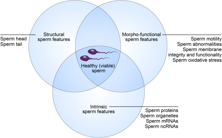
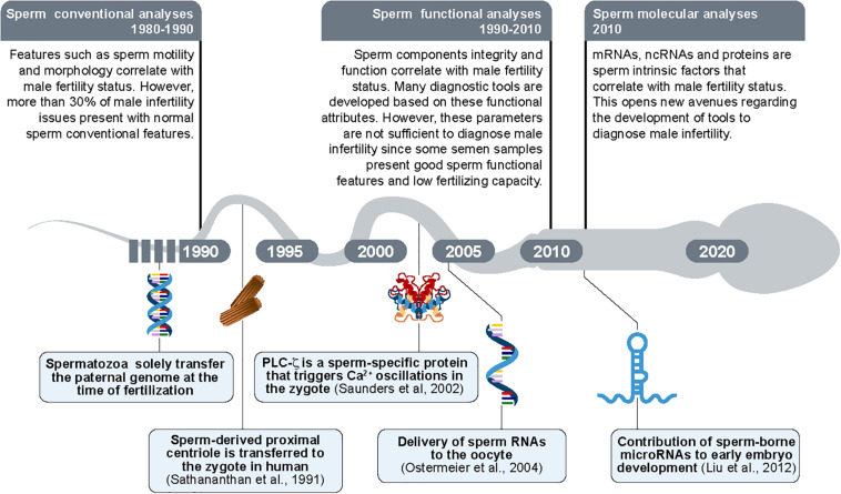
In addition to the paternal genome, spermatozoa carry several intrinsic factors, including organelles (e.g., centrioles and mitochondria) and molecules (e.g., proteins and RNAs), which are involved in important steps of reproductive biology such as spermatogenesis, sperm maturation, oocyte fertilization and embryo development. These factors constitute potential biomarkers of "viable sperm" and male fertility status and may become major assets for diagnosing instances of idiopathic male infertility in both humans and livestock animals. A better understanding of the mechanism of action of these sperm intrinsic factors in the regulation of reproductive and developmental processes still presents a major challenge that must be addressed. This review assembles the main data regarding morpho-functional and intrinsic sperm features that are associated with male infertility, with a particular focus on microRNA (miRNA) molecules.

Keywords: cattle; diagnosis; human; infertility; intrinsic factors; ncRNAs; semen; spermatozoa.


Figures

FIGURE 1
FIGURE 1
Concept of “healthy (viable) sperm.” Male fertility potential relies on structural, morpho-functional, and intrinsic sperm features that shape equally the concept of “healthy (viable) sperm.”
FIGURE 2
FIGURE 2
Structural sperm features. Spermatozoa are composed of two main parts: head and tail (or flagellum). The sperm head is constituted basically by the acrosome and nucleus. The sperm tail includes: the neck that contains mainly the proximal centriole; the midpiece which is composed by mitochondria, outer dense fibers (ODF) and axoneme; principal piece containing the fibrous sheath and axoneme; and terminal piece.
FIGURE 3
FIGURE 3
Morpho-functional sperm features. Schematic figure representing the spermatozoa with satisfactory (left) and unsatisfactory (right) morpho-functional features. Sperm acrosome membrane integrity, sperm plasma membrane integrity, sperm DNA integrity, low quantity of ROS, sperm mitochondrial membrane high activity, high sperm motility and normal sperm morphology characterize the satisfactory morpho-functional sperm features. Sperm acrosome membrane damage, sperm plasma membrane damage, sperm DNA fragmentation, high quantity of ROS, sperm mitochondrial membrane low activity, low sperm motility and abnormal sperm morphology characterize the unsatisfactory morpho-functional sperm features.
FIGURE 4
FIGURE 4
Intrinsic sperm features. Spermatozoon contributes early embryo development during mature oocyte fertilization, zygote formation and embryo cleavage potentially with intrinsic sperm features such as: sperm proteins (e.g., PLCζ, to promote oocyte activation); sperm RNAs and microRNAs (miRNAs); sperm DNA, to generate male pronucleus; sperm centrioles, to form sperm aster; and may contribute sperm mitochondria, promoting mtDNA heteroplasmy.
FIGURE 5
FIGURE 5
Schematic figure demonstrating the contribution of microRNAs (miRNAs) in the reproductive events. The sperm-related-miRNAs molecules display functions in spermatogenesis (testis), sperm maturation (epididymis caput, corpus, and cauda), sperm and seminal plasma interaction (e.g., epididymosomes) as well as modulating early embryo development.
FIGURE 6
FIGURE 6
Timeline of the new findings regarding sperm morpho-functional and intrinsic features. The paternal DNA was considered as the sole intrinsic sperm feature transferred from spermatozoon to oocyte until 1990s. In parallel, the sperm evaluation was limited to sperm conventional analyses (sperm motility and sperm morphology/abnormalities). Proximal centrioles were then shown to be transmitted by sperm to the oocytes during fertilization for the first time in 1991 (Sathananthan et al., 1991). The sperm-borne PLCζ protein was shown as a promotor of oocyte activation in 2002 (Saunders et al., 2002). The delivery of RNAs molecules was then revealed as transferred from sperm to the oocyte in 2004 (Ostermeier et al., 2004). In parallel, the sperm evaluation was updated to sperm functional analyses (e.g., sperm plasma membrane integrity). In 2010s, small sperm-borne RNAs molecules (microRNAs/miRNAs) were shown as important to early embryo development (Liu et al., 2012) potentially constituting a new group of sperm analyses composed by evaluation of molecular targets.

References

    1. Aarabi M., Balakier H., Bashar S., Moskovtsev S. I., Sutovsky P., Librach C. L., et al. (2014). Sperm content of postacrosomal WW binding protein is related to fertilization outcomes in patients undergoing assisted reproductive technology. Fertil. Steril. 102 440–447. 10.1016/j.fertnstert.2014.05.003 - DOI - PubMed
    1. Abu-Halima M., Hammadeh M., Schmitt J., Leidinger P., Keller A., Meese E., et al. (2013). Altered microRNA expression profiles of human spermatozoa in patients with different spermatogenic impairments. Fertil. Steril. 99 1249.e16–1255.e16. 10.1016/j.fertnstert.2012.11.054 - DOI - PubMed
    1. Agarwal A., Mulgund A., Hamada A., Chyatte M. R. (2015). A unique view on male infertility around the globe. Reprod. Biol. Endocrinol. 13 1–9. 10.1186/s12958-015-0032-1 - DOI - PMC - PubMed
    1. Aitken R. J., Wingate J. K., De Iuliis G. N., McLaughlin E. A. (2007). Analysis of lipid peroxidation in human spermatozoa using BODIPY C11. MHR Basic Sci. Reprod. Med. 13 203–211. 10.1093/molehr/gal119 - DOI - PubMed
    1. Alves M. B. R., de Arruda R. P., De Bem T. H. C., Florez-Rodriguez S. A., Sá Filho M. F., de Belleannée C., et al. (2019). Sperm-borne miR-216b modulates cell proliferation during early embryo development via K-RAS. Sci. Rep. 9 1–14. 10.1038/s41598-019-46775-8 - DOI - PMC - PubMed